What Does The Circulatory System Do
The circulatory system is made up of blood vessels that carry blood away from and towards the heart. Arteries carry blood away from the heart and veins carry blood back to the heart.
The circulatory system carries oxygen, nutrients, and to cells, and removes waste products, like carbon dioxide. These roadways travel in one direction only, to keep things going where they should.
How The Heart Works
The heart is an organ, about the size of a fist. It is made of muscle and pumps blood through the body. Blood is carried through the body in blood vessels, or tubes, called arteries and veins. The process of moving blood through the body is called circulation. Together, the heart and vessels make up the cardiovascular system.
Blood Flow To The Heart
After the blood has passed through the capillary beds, it enters the venules, veins, and finally the two main venae cavae that take blood back to the heart. The flow rate increases again, but is still much slower than the initial rate in the aorta. Blood primarily moves in the veins by the rhythmic movement of smooth muscle in the vessel wall and by the action of the skeletal muscle as the body moves. Because most veins must move blood against the pull of gravity, blood is prevented from flowing backward in the veins by one-way valves. Thus, because skeletal muscle contraction aids in venous blood flow, it is important to get up and move frequently after long periods of sitting so that blood will not pool in the extremities.
- This page has no tags.
You May Like: Atropine Arrhythmia
What Are The Coronary Arteries Of The Heart
Like all organs, your heart is made of tissue that requires a supply of oxygen and nutrients. Although its chambers are full of blood, the heart receives no nourishment from this blood. The heart receives its own supply of blood from a network of arteries, called the coronary arteries.
Two major coronary arteries branch off from the aorta near the point where the aorta and the left ventricle meet:
- Right coronary artery supplies the right atrium and right ventricle with blood. It branches into the posterior descending artery, which supplies the bottom portion of the left ventricle and back of the septum with blood.
- Left main coronary artery branches into the circumflex artery and the left anterior descending artery. The circumflex artery supplies blood to the left atrium, side and back of the left ventricle, and the left anterior descending artery supplies the front and bottom of the left ventricle and the front of the septum with blood.
These arteries and their branches supply all parts of the heart muscle with blood.
Coronary artery disease occurs when plaque builds up in the coronary arteries and prevents the heart from getting the enriched blood it needs. If this happens, a network of tiny blood vessels in the heart that aren’t usually open called collateral vessels may enlarge and become active. This allows blood to flow around the blocked artery to the heart muscle, protecting the heart tissue from injury.
About The Heart And Circulatory System

The circulatory system is composed of the heart and blood vessels, including arteries, veins, and capillaries. Our bodies actually have two circulatory systems: The pulmonary circulation is a short loop from the heart to the lungs and back again, and the systemic circulation sends blood from the heart to all the other parts of our bodies and back again.
The heart is the key organ in the circulatory system. As a hollow, muscular pump, its main function is to propel blood throughout the body. It usually beats from 60 to 100 times per minute, but can go much faster when necessary. It beats about 100,000 times a day, more than 30 million times per year, and about 2.5 billion times in a 70-year lifetime.
The heart gets messages from the body that tell it when to pump more or less blood depending on an individuals needs. When were sleeping, it pumps just enough to provide for the lower amounts of oxygen needed by our bodies at rest. When were exercising or frightened, the heart pumps faster to increase the delivery of oxygen.
The heart has four chambers that are enclosed by thick, muscular walls. It lies between the lungs and just to the left of the middle of the chest cavity. The bottom part of the heart is divided into two chambers called the right and left ventricles, which pump blood out of the heart. A wall called the interventricular septum divides the ventricles.
Arterial walls have three layers:
You May Like: How Does Blood Move Through The Heart
Supplying Oxygen To The Hearts Muscle
Like other muscles in the body, your heart needs blood to get oxygen and nutrients. Yourcoronary arteries supply blood to your heart. These arteries branch off from the aorta so that oxygen-rich blood is delivered to your heart as well as the rest of your body.
- The left coronary artery delivers blood to the left side of your heart, including your left atrium and ventricle and the septum between the ventricles.
- The circumflex artery branches off from the left coronary artery to supply blood to part of the left ventricle.
- The left anterior descending artery also branches from the left coronary artery and provides blood to parts of both the right and left ventricles.
- The right coronary artery provides blood to the right atrium and parts of both ventricles.
- The marginal arteries branch from the right coronary artery and provide blood to the surface of the right atrium.
- The posterior descending artery also branches from the right coronary artery and provides blood to the bottom of both ventricles.
Arteries supplying oxygen to the body. The coronary arteries branch off the aorta and supply the heart muscle with oxygen and nutrients. At the top of your aorta, arteries branch off to carry blood to your head and arms. Arteries branching from the middle and lower parts of your aorta supply blood to the rest of your body.
Some conditions can affect normal blood flow through these heart arteries. Examples include:
- The small cardiac vein.
How Does Blood Flow To Lungs
Through the pulmonic valves, the blood can enter into your pulmonary. Then the blood starts the travel of pulmonary circulation.
From the right side of the heart, blood flows past the pulmonic valve to enter your lungs through the pulmonary circulation. It travels into the lungs through your pulmonary artery and passes into the tiny capillaries in the lungs. The capillaries have very thin walls that allow oxygen from the air sacs in your lungs to be absorbed, at the same time allowing carbon dioxide, a metabolic waste product, to pass from your blood into these air sacs. CO2 is then eliminated whenever you exhale. The oxygenated blood is sent back to the left side of the heart through your pulmonary veins.
You May Like: Does Tylenol Elevate Blood Pressure
What Does The Heart Look Like And How Does It Work
- The heart is an amazing organ. It starts beating about 22 days after conception and continuously pumps oxygenated red blood cells and nutrient-rich blood and other compounds like platelets throughout your body to sustain the life of your organs.
- Its pumping power also pushes blood through organs like the lungs to remove waste products like CO2.
- This fist-sized powerhouse beats about 100,000 times per day, pumping five or six quarts of blood each minute, or about 2,000 gallons per day.
- In general, if the heart stops beating, in about 4-6 minutes of no blood flow, brain cells begin to die and after 10 minutes of no blood flow, the brain cells will cease to function and effectively be dead. There are few exceptions to the above.
- The heart works by a regulated series of events that cause this muscular organ to contract and then relax .
- The normal heart has 4 chambers that undergo the squeeze and relax cycle at specific time intervals that are regulated by a normal sequence of electrical signals that arise from specialized tissue.
- In addition, the normal sequence of electrical signals can be sped up or slowed down depending on the needs of the individual, for example, the heart will automatically speed up electrical signals to respond to a person running and will automatically slow down when a person takes a nap.
The Left Side Of The Heart
Oxygen-rich blood from the lungs passes through the pulmonary veins . It enters the left atrium and is pumped into the left ventricle. From the left ventricle, the blood is pumped to the rest of the body through the aorta.
Like all of the organs, the heart needs blood rich with oxygen. This oxygen is supplied through the coronary arteries as its pumped out of the hearts left ventricle.
The coronary arteries are located on the hearts surface at the beginning of the aorta. The coronary arteries carry oxygen-rich blood to all parts of the heart.
You May Like: What Are The Early Signs Of Congestive Heart Failure
How Does Blood Travel Tbrough The Heart
It is pumped through. The heart is a muscle that rhymically and regulary contracts. It has chambers which hold the blood, and one-way valves which control the flow of blood out from the contracting chamber.
Different creatures can have different heart structures.
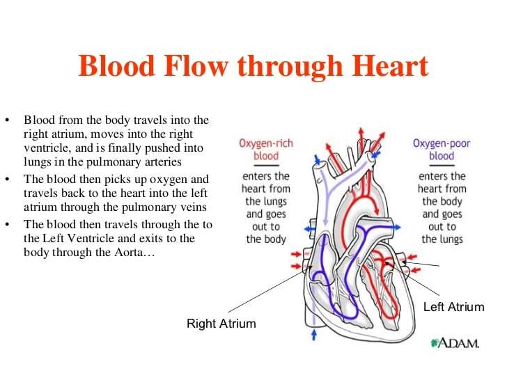
The human heart is a double pump, and has four chambers through which the blood flows, de-oxygenated blood flowing IN from the body into the right atrium, and pumped down into the right ventricle, then OUT to the lungs, via the pulmonary arteries, for ‘carbon dioxode for oxygen’ gas exchange.
The oxygenated blood then comes back into the heart, via the pulmonary veins, into the left atrium, and is pumped down into the left ventricle, from where it is pumped, via the massive aorta, to the main arteries of the body.
How Does The Heart Beat
The ventricles and atria work together and alternately relax and contract for making the blood flow through the heart. Your hearts electrical system provides them the power for accomplishing this goal.
The impulse begins in a little group of specialized and efficient cells known as the sinoatrial node situated in your right atrium. The SA node is also referred to as your hearts pacemaker. The electrical impulse spreads via the walls of your atria, stimulating them to instantly contract.
A group of cells lying in your hearts center between your ventricles and atria known as the atrioventricular node works like a door that reduces the speed of the electrical impulse before it moves to the ventricles. This slight delay gives your atria enough time to quickly contract prior to the contraction of the ventricles.
The His-Purkinje network refers to a path of numerous fibers that send that electrical impulse to your ventricles muscular walls, triggering them to quickly contract.
The Heart Rate
When you are resting, your heart beats about 50 to around 99 times per minute. Medications, emotions, fever and exercise can make your heart beat faster. At times, the heart can beat more than 100 beats/ minute.
Don’t Miss: Ibs And Heart Palpitations
S Of Blood Flow Through The Heart
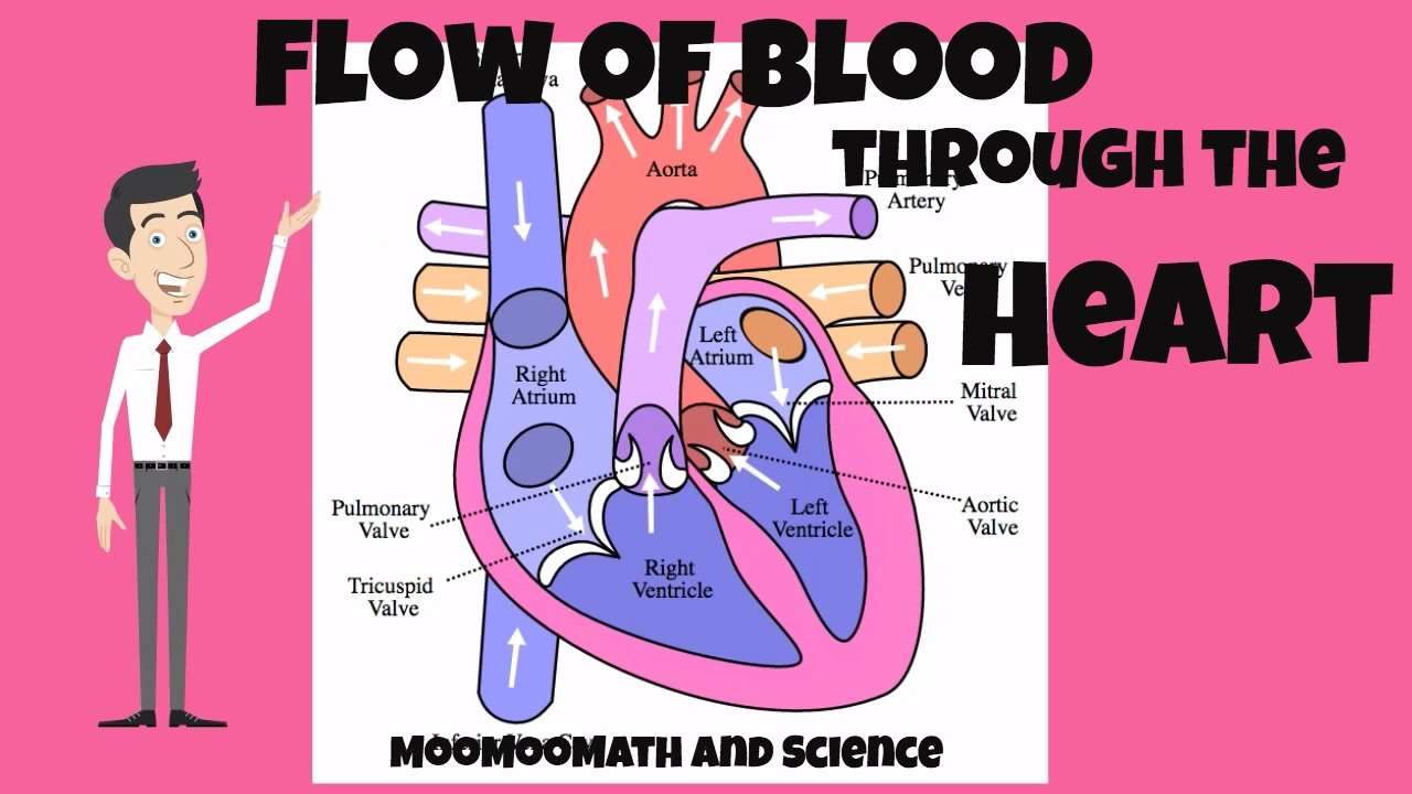
In summary from the video, in 14 steps, blood flows through the heart in the following order: 1) body > 2) inferior/superior vena cava > 3) right atrium > 4) tricuspid valve > 5) right ventricle > 6) pulmonary arteries > 7) lungs > 8) pulmonary veins > 9) left atrium > 10) mitral or bicuspid valve > 11) left ventricle > 12) aortic valve > 13) aorta > 14) body.
Where Is Your Heart And What Does It Look Like

The heart is located under the rib cage, to the left of your breastbone and between your lungs.
Looking at the outside of the heart, you can see that the heart is made of muscle. The strong muscular walls contract , pumping blood to the arteries. The major blood vessels connected to your heart are the aorta, the superior vena cava, the inferior vena cava, the pulmonary artery , the pulmonary veins , and the coronary arteries .
On the inside, the heart is a four-chambered, hollow organ. It is divided into the left and right side by a wall called the septum. The right and left sides of the heart are further divided into two top chambers called the atria, which receive blood from the veins, and two bottom chambers called ventricles, which pump blood into the arteries.
The atria and ventricles work together, contracting and relaxing to pump blood out of the heart. As blood leaves each chamber of the heart, it passes through a valve. There are four heart valves within the heart:
- Mitral valve
- Aortic valve
- Pulmonic valve
The tricuspid and mitral valves lie between the atria and ventricles. The aortic and pulmonic valves lie between the ventricles and the major blood vessels leaving the heart.
The heart valves work the same way as one-way valves in the plumbing of your home. They prevent blood from flowing in the wrong direction.
Recommended Reading: Low Blood Pressure Heart Failure Elderly
Anatomy Of Your Heart
Your heart is in the center of your chest, near your lungs. It has four hollow heart chambers surrounded by muscle and other heart tissue. The chambers are separated by heart valves, which make sure that the blood keeps flowing in the right direction. Read more about heart valves in Blood Flow.
Anatomy of the interior of the heart. This image shows the four chambers of the heart and the direction that blood flows through the heart. Oxygen-poor blood, shown in blue-purple, flows into the heart and is pumped out to the lungs. Then oxygen-rich blood, shown in red, is pumped out to the rest of the body, with the help of the heart valves.
Left Side Of The Heart
- The pulmonary veins empty oxygen-rich blood from the lungs into the left atrium of the heart.
- As the atrium contracts, blood flows from your left atrium into your left ventricle through the open mitral valve.
- When the ventricle is full, the mitral valve shuts. This prevents blood from flowing backward into the atrium while the ventricle contracts.
- As the ventricle contracts, blood leaves the heart through the aortic valve, into the aorta and to the body.
Don’t Miss: Esophagus Palpitations
Blood Flow Away From The Heart
With each rhythmic pump of the heart, blood is pushed under high pressure and velocity away from the heart, initially along the main artery, the aorta. In the aorta, the blood travels at 30 cm/sec. From the aorta, blood flows into the arteries and arterioles and, ultimately, to the capillary beds. As it reaches the capillary beds, the rate of flow is dramatically slower than the rate of flow in the aorta. While the diameter of each individual arteriole and capillary is far narrower than the diameter of the aorta, the rate is actually slower due to the overall diameter of all the combined capillaries being far greater than the diameter of the individual aorta.
View of the heartPrecapillary sphincters
Blood Flow Of The Heart Review
Lets now use the 2×2 table we made in the anatomy of the heart post, and this will give us another way to visualize the blood flow through the heart.
Right Side
First, we have the SVC and IVC that carry deoxygenated venous blood from the rest of the body to the right atrium.
Blood will then flow from the right atrium, through the tricuspid valve, and enter the right ventricle.
The deoxygenated blood will then exit the right ventricle, travel through the pulmonary valve, and enter the main pulmonary artery to ultimately be delivered to the lungs to become oxygenated.
Left Side
The oxygenated blood will then travel from the lungs to the left atrium via the pulmonary veins.
Blood will then flow from the left atrium, through the mitral valve, and enter the left ventricle.
The oxygenated blood will then exit the left ventricle, travel through the aortic valve, and enter the aorta to be delivered to the rest of the body.
Diagram: Blood flow through the heart, cardiac circulation pathway steps, and cardiac anatomy and structures. Blue arrows Red arrows .
Now that we have a good understanding of the blood flow through the heart using the cartoon diagrams, we can apply it to a more realistic image of the heart.
The blue arrows represent the flow of deoxygenated blood through the right side of the heart.
The red arrows represent the flow of oxygenated blood through the left side of the heart.
Don’t Miss: Heart Attack Grill Blair River
Blood Flow Positive And Negative Effects
A healthy heart normally beats anywhere from 60 to 70 times per minute when you’re at rest. This rate can be higher or lower depending on your health and physical fitness athletes generally have a lower resting heart rate, for example.
Your heart rate rises with physical activity, as your muscles consume oxygen while they work. The heart works harder to bring oxygenated blood where it is needed.
Disrupted or irregular heartbeats can affect blood flow through the heart. This can happen in multiple ways:
- Electrical impulses that regulate your heartbeat are impacted, causing an arrythmia, or irregular heartbeat. Atrial fibrillation is a common form of this.
- Conduction disorders, or heart blocks, affect the cardiac conduction system, which regulates how electrical impulses move through the heart. The type of blockan atrioventricular block or bundle branch blockdepends on where it occurs in the conduction system.
- Damaged or diseased valves can become ineffective or leak blood in the wrong direction.
- A blocked blood vessel, which can happen gradually or suddenly, can disrupt blood flow, such as during a heart attack.
If you experience an irregular heartbeat or cardiac symptoms like chest pain and shortness of breath, seek medical help immediately.
