How Does Your Heart Work
Your heart is made up of 2 pumps. The pump on the right hand side receives blood that has already delivered its oxygen round the body and sends this blood to the lungs to pick up more oxygen .
The pump on the left hand side receives oxygen-rich blood and then pumps it out into the arteries to deliver its oxygen around the body.
What Does The Circulatory System Do
The circulatory system is made up of blood vessels that carry blood away from and towards the heart. Arteries carry blood away from the heart and veins carry blood back to the heart.
The circulatory system carries oxygen, nutrients, and to cells, and removes waste products, like carbon dioxide. These roadways travel in one direction only, to keep things going where they should.
How Can I Help Keep My Child’s Heart Healthy
To help keep your child’s heart healthy:
- Encourage plenty of exercise.
- Help your child reach and keep a healthy weight.
- Go for regular medical checkups.
- Tell the doctor about any family history of heart problems.
Let the doctor know if your child has any chest pain, trouble breathing, or dizzy or fainting spells or if your child feels like the heart sometimes goes really fast or skips a beat.
Recommended Reading: Can Flonase Cause Heart Palpitations
What Are The Coronary Arteries Of The Heart
Like all organs, your heart is made of tissue that requires a supply of oxygen and nutrients. Although its chambers are full of blood, the heart receives no nourishment from this blood. The heart receives its own supply of blood from a network of arteries, called the coronary arteries.
Two major coronary arteries branch off from the aorta near the point where the aorta and the left ventricle meet:
- Right coronary artery supplies the right atrium and right ventricle with blood. It branches into the posterior descending artery, which supplies the bottom portion of the left ventricle and back of the septum with blood.
- Left main coronary artery branches into the circumflex artery and the left anterior descending artery. The circumflex artery supplies blood to the left atrium, side and back of the left ventricle, and the left anterior descending artery supplies the front and bottom of the left ventricle and the front of the septum with blood.
These arteries and their branches supply all parts of the heart muscle with blood.
Coronary artery disease occurs when plaque builds up in the coronary arteries and prevents the heart from getting the enriched blood it needs. If this happens, a network of tiny blood vessels in the heart that aren’t usually open called collateral vessels may enlarge and become active. This allows blood to flow around the blocked artery to the heart muscle, protecting the heart tissue from injury.
How Does Blood Flow Through Your Lungs

Once blood travels through the pulmonic valve, it enters your lungs. This is called the pulmonary circulation. From your pulmonic valve, blood travels to the pulmonary artery to tiny capillary vessels in the lungs. Here, oxygen travels from the tiny air sacs in the lungs, through the walls of the capillaries, into the blood. At the same time, carbon dioxide, a waste product of metabolism, passes from the blood into the air sacs. Carbon dioxide leaves the body when you exhale. Once the blood is purified and oxygenated, it travels back to the left atrium through the pulmonary veins.
You May Like: Does Tylenol Increase Heart Rate
Left Side Of The Heart
- The pulmonary veins empty oxygen-rich blood from the lungs into the left atrium of the heart.
- As the atrium contracts, blood flows from your left atrium into your left ventricle through the open mitral valve.
- When the ventricle is full, the mitral valve shuts. This prevents blood from flowing backward into the atrium while the ventricle contracts.
- As the ventricle contracts, blood leaves the heart through the aortic valve, into the aorta and to the body.
Function Of The Pericardium
The pericardium is important because it protects the heart from trauma, shock, stress, and even infections from the nearby lungs. It supports the heart and anchors it to the medastinum so it doesnt move within the body. The pericardium lubricates the heart and prevents it from becoming too large if blood volume is overloaded .
Despite these functions, the pericardium is still vulnerable to problems of its own. Pericarditis is the term for inflammation in the pericardium, typically due to infection. Pericarditis is often a severe disease because it can constrict and apply pressure on the heart and work against its normal function. Pericarditis comes in many types depending on which tissue layer is infected.
Donât Miss: Does Benadryl Increase Your Heart Rate
You May Like: Flonase And Heart Palpitations
The Systemic Loop Goes All Over The Body
In the systemic loop, oxygenated blood is pumped from the left ventricle of the heart through the aorta, the largest artery in the body. The blood moves from the aorta through the systemic arteries, then to arterioles and capillary beds that supply body tissues. Here, oxygen and nutrients are released and carbon dioxide and other waste substances are absorbed. Deoxygenated blood then moves from the capillary beds through venules into the systemic veins. The systemic veins feed into the inferior and superior venae cavae, the largest veins in the body. The venae cavae flow deoxygenated blood to the right atrium of the heart.
Recommended Reading: Does Benadryl Lower Heart Rate
Heart Contraction And Blood Flow
Almost everyone has heard the real or recorded sound of a heartbeat. When the heart beats, it makes a lub-DUB sound. Between the time you hear lub and DUB, blood is pumped through the heart and circulatory system.
A heartbeat may seem like a simple event repeated over and over. A heartbeat actually is a complicated series of very precise and coordinated events that take place inside and around the heart.
Each side of the heart uses an inlet valve to help move blood between the atrium and ventricle.
The tricuspid valve does this between the right atrium and ventricle. The mitral valve does this between the left atrium and ventricle. The lub is the sound of the mitral and tricuspid valves closing.
Each of the hearts ventricles has an outlet valve. The right ventricle uses the pulmonary valve to help move blood into the pulmonary arteries. The left ventricle uses the aortic valve to do the same for the aorta. The DUB is the sound of the aortic and pulmonary valves closing.
Each heartbeat has two basic parts: diastole and atrial and ventricular systole . During diastole, the atria and ventricles of the heart relax and begin to fill with blood.
At the end of diastole, the hearts atria contract and pump blood into the ventricles. The atria then begin to relax. Next, the hearts ventricles contract and pump blood out of the heart.
Recommended Reading: How To Calculate Target Heart Rate Zone
When Should I Talk To A Doctor
You should call your healthcare provider if you experience:
- Bluish lips or skin color.
- Swollen ankles, feet or abdomen.
A note from Cleveland Clinic
Your pulmonary arteries play an important role getting carbon dioxide out of your blood and oxygen back into it. Many conditions that affect the pulmonary arteries and pulmonary blood circulation are congenital or present at birth. But coronary artery disease and other heart disease can damage the pulmonary arteries. Depending on the heart problem, you may need surgery or other treatments to improve blood flow and oxygenation. Your healthcare provider can offer suggestions on ways to improve your heart health.
Last reviewed by a Cleveland Clinic medical professional on 03/10/2021.
References
- Adult Congenital Heart Association. . Accessed 3/10/2021.Pulmonary Hypertension
- American Heart Association. Problem: . Accessed 3/10/2021.Pulmonary Valve Regurgitation
- Castaner E, Gallardo X, Rimola J, et al. Radiologic overview. RadioGraphics. Accessed 3/10/2021.Congenital and acquired pulmonary artery anomalies in the adult:
- Kreibich M, Siepe M, Kroll J, et al. Circulation. 2015 131:310-16. Accessed 3/10/2021.Aneurysms of the pulmonary artery.
- Tucker WB, Weber C, Burns B. . StatPearls . 2020. Accessed 3/10/2021.Anatomy, thorax, heart pulmonary arteries
Cleveland Clinic is a non-profit academic medical center. Advertising on our site helps support our mission. We do not endorse non-Cleveland Clinic products or services.Policy
The Cardiac Cycle Includes All Blood Flow Events The Heart Accomplishes In One Complete Heartbeat
The muscular wall of the heart powers contraction and dilation. Each contraction and relaxation is a heartbeat. Ventricular contractions, called systole, force blood out of the heart through the pulmonary and aortic valves. Diastole occurs when blood flows from the atria to fill the ventricles.
Read Also: Does Benadryl Lower Heart Rate
You May Like: Reflux And Palpitations
Which Heart Chamber Sends Deoxygenated Blood To The Lungs
4.4/5right ventricleright atriumleft ventricleleft atrium
Deoxygenated blood leaves the heart, goes to the lungs, and then re-enters the heart Deoxygenated blood leaves through the right ventricle through the pulmonary artery. From the right atrium, the blood is pumped through the tricuspid valve , into the right ventricle.
One may also ask, which heart chamber receives blood from the pulmonary veins quizlet? left atrium
Also asked, which part of the heart pumps blood to the lungs?
The right side of the heart pumps blood to the lungs to pick up oxygen. The left side of the heart receives the oxygen-rich blood from the lungs and pumps it to the body.
How does oxygenated and deoxygenated blood flow through the heart?
Systemic circulation carries oxygenated blood from the left ventricle, through the arteries, to the capillaries in the tissues of the body. From the tissue capillaries, the deoxygenated blood returns through a system of veins to the right atrium of the heart.
How The Heart Works

The heart is a large, muscular organ that pumps blood filled with oxygen and nutrients through the blood vessels to the body tissues. It’s made up of:
-
4 chambers. The 2 upper chambers are the atria. They receive and collect blood. The 2 lower chambers are the ventricles. They pump blood to other parts of your body. Here is the process:
-
The right atrium receives blood from the body. This blood is low in oxygen. This is the blood from the veins.
-
The right ventricle pumps the blood from the right atrium into the lungs to pick up oxygen and remove carbon dioxide.
-
The left atrium receives blood from the lungs. This blood is rich in oxygen.
-
The left ventricle pumps the blood from the left atrium out to the body, supplying all organs with oxygen-rich blood.
4 valves. The 4 valves are the aortic, pulmonary, mitral, and tricuspid valves. They let blood flow forward and prevent the backward flow.
Blood vessels. These bring blood to the lungs, where oxygen enters the bloodstream, and then to the body:
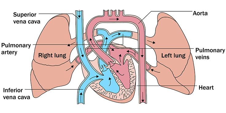
The inferior and superior vena cava bring oxygen-poor blood from the body into the right atrium.
The pulmonary artery channels oxygen-poor blood from the right ventricle into the lungs, where oxygen enters the bloodstream.
The pulmonary veins bring oxygen-rich blood to the left atrium.
The aorta channels oxygen-rich blood to the body from the left ventricle.
An electrical system that stimulates contraction of the heart muscle.
Also Check: Can Ibs Cause Heart Palpitations
The Pulmonary Loop Only Transports Blood Between The Heart And Lungs
In the pulmonary loop, deoxygenated blood exits the right ventricle of the heart and passes through the pulmonary trunk. The pulmonary trunk splits into the right and left pulmonary arteries. These arteries transport the deoxygenated blood to arterioles and capillary beds in the lungs. There, carbon dioxide is released and oxygen is absorbed. Oxygenated blood then passes from the capillary beds through venules into the pulmonary veins. The pulmonary veins transport it to the left atrium of the heart. The pulmonary arteries are the only arteries that carry deoxygenated blood, and the pulmonary veins are the only veins that carry oxygenated blood.
The Interior Of The Heart
Below is a picture of the inside of a normal, healthy, human heart.
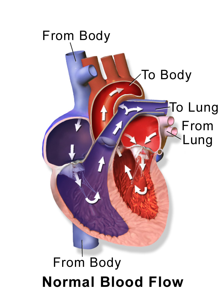
The illustration shows a cross-section of a healthy heart and its inside structures. The blue arrow shows the direction in which low-oxygen blood flows from the body to the lungs. The red arrow shows the direction in which oxygen-rich blood flows from the lungs to the rest of the body.
Read Also: What Heart Chamber Pushes Blood Through The Aortic Semilunar Valve
How The Normal Heart Works
The heart is a large muscular organ with the very important job of circulating blood through the blood vessels to the body. Located in the center of the chest, the heart is the hardest working muscle in the human body always working, even while we are sleeping. The heart and blood vessels together make up the body’s cardiovascular system and are vital to supplying the body with the necessary oxygen and nutrients needed to survive. When you breathe, your lungs take in oxygen. The heart pumps blood to the lungs to pick up oxygen, and then it pumps blood through the body to deliver that oxygen.
The animations below show how a normal heart pumps blood. They also explain the changes that happen to a normal heart right after the fetus is born.
Each Heart Beat Is A Squeeze Of Two Chambers Called Ventricles
The ventricles are the two lower chambers of the heart. Blood empties into each ventricle from the atrium above, and then shoots out to where it needs to go. The right ventricle receives deoxygenated blood from the right atrium, then pumps the blood along to the lungs to get oxygen. The left ventricle receives oxygenated blood from the left atrium, then sends it on to the aorta. The aorta branches into the systemic arterial network that supplies all of the body.
Also Check: Can Antihistamines Cause Heart Palpitations
What Are The Parts Of The Circulatory System
Two pathways come from the heart:
- The pulmonary circulation is a short loop from the heart to the lungs and back again.
- The systemic circulation carries blood from the heart to all the other parts of the body and back again.
In pulmonary circulation:
- The pulmonary artery is a big artery that comes from the heart. It splits into two main branches, and brings blood from the heart to the lungs. At the lungs, the blood picks up oxygen and drops off carbon dioxide. The blood then returns to the heart through the pulmonary veins.
In systemic circulation:
Left Ventricle Sends Oxygen
The left ventricle relaxes and fills up with blood before squeezing and pumping the oxygen-rich blood through the aortic valve into the aorta the main artery that carries blood to your body. The muscle wall of the left ventricle is very thick because it has to pump blood around the whole body.
Don’t Miss: Can Ibs Cause Heart Palpitations
The Heart’s Control System
A heartbeat is caused by an electrical impulse traveling through the heart. The heart’s built-in electrical system controls the speed of its pumping. The electrical impulse originates in the sinus node which functions as the heart’s natural pacemaker. The sinus node is most often located in the top of the right atrium. The electrical signals travel through the heart tissue causing the atria and ventricles to contract and relax and the blood to be pumped to the body.
Anatomy Of Your Heart

Your heart is in the center of your chest, near your lungs. It has four hollow heart chambers surrounded by muscle and other heart tissue. The chambers are separated by heart valves, which make sure that the blood keeps flowing in the right direction. Read more about heart valves in Blood Flow.
Anatomy of the interior of the heart. This image shows the four chambers of the heart and the direction that blood flows through the heart. Oxygen-poor blood, shown in blue-purple, flows into the heart and is pumped out to the lungs. Then oxygen-rich blood, shown in red, is pumped out to the rest of the body, with the help of the heart valves.
Also Check: How To Find Thrz
Heart Diagram Parts Location And Size
Location and size of the heart
Normal heart anatomy and physiology
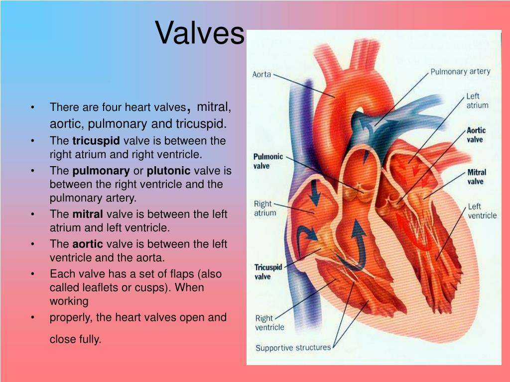
Normal heart anatomy and physiology need the atria and ventricles to work sequentially, contracting and relaxing to pump blood out of the heart and then to let the chambers refill. When blood leaves each chamber of the heart, it passes through a valve that is designed to prevent backflow of blood. There are four heart valves within the heart:
- Mitral valve between the left atrium and left ventricle
- Tricuspid valve between the right atrium and right ventricle
- Aortic valve between the left ventricle and aorta
- Pulmonic valve between the right ventricle and pulmonary artery
How the heart valves work
Electrical Impulses Keep The Beat
The heart’s four chambers pump in an organized manner with the help of electrical impulses that originate in the sinoatrial node . Situated on the wall of the right atrium, this small cluster of specialized cells is the heart’s natural pacemaker, initiating electrical impulses at a normal rate.
The impulse spreads through the walls of the right and left atria, causing them to contract, forcing blood into the ventricles. The impulse then reaches the atrioventricular node, which acts as an electrical bridge for impulses to travel from the atria to the ventricles. From there, a pathway of fibers carries the impulse into the ventricles, which contract and force blood out of the heart.
Read Also: Is Tylenol Bad For Your Heart
The Valves Are Like Doors To The Chambers Of The Heart
Four valves regulate and support the flow of blood through and out of the heart. The blood can only flow one waylike a car that must always be kept in drive. Each valve is formed by a group of folds, or cusps, that open and close as the heart contracts and dilates. There are two atrioventricular valves, located between the atrium and the ventricle on either side of the heart: The tricuspid valve on the right has three cusps, the mitral valve on the left has two. The other two valves regulate blood flow out of the heart. The aortic valve manages blood flow from the left ventricle into the aorta. The pulmonary valve manages blood flow out of the right ventricle through the pulmonary trunk into the pulmonary arteries.
Also Check: Does Tylenol Increase Heart Rate
