What Is Lad And Rca Disease
LAD: left anterior descending coronary artery LCx: left circumflex coronary artery RCA: right coronary artery SCA: single coronary artery. This case demonstrates a rare congenital coronary artery anomaly found incidentally with a presentation of atypical angina and three-vessel coronary artery disease .
Improving Health With Current Research
Learn about the following ways the NHLBI continues to translate current research and science into improved health for people who have heart conditions. Research on this topic is part of the NHLBI’s broader commitment to advancing heart and vascular disease scientific discovery.
Learn about some of the pioneering research contributions we have made over the years that have improved clinical care.
Anatomy Of The Heart And Blood Vessels
Reviewed byDr Jacqueline Payne
The heart is a muscular pump that pushes blood through blood vessels around the body. The heart beats continuously, pumping the equivalent of more than 14,000 litres of blood every day through five main types of blood vessels: arteries, arterioles, capillaries, venules and veins.
Also Check: Tylenol And Heart Rate
About Blood Supply Of Heart
The heart is a major organ of the human body which is responsible for the circulation of the blood. Blood circulation in the body ensures that every cell of the organ receives nutrients and oxygen supply to perform its metabolism. To understand the blood supply of the heart it is important to understand the location of the heart, anatomy of the heart, the vasculature of the nerve which answers questions like which artery supplies blood to the heart. This article dwells with the intricacies of blood supply of heart anatomy and functioning, which arteries supply blood to heart, the circulation system of the heart known as coronary circulation.
How The Heart Works

The heart is a large, muscular organ that pumps blood filled with oxygen and nutrients through the blood vessels to the body tissues. It’s made up of:
-
4 chambers. The 2 upper chambers are the atria. They receive and collect blood. The 2 lower chambers are the ventricles. They pump blood to other parts of your body. Here is the process:
-
The right atrium receives blood from the body. This blood is low in oxygen. This is the blood from the veins.
-
The right ventricle pumps the blood from the right atrium into the lungs to pick up oxygen and remove carbon dioxide.
-
The left atrium receives blood from the lungs. This blood is rich in oxygen.
-
The left ventricle pumps the blood from the left atrium out to the body, supplying all organs with oxygen-rich blood.
4 valves. The 4 valves are the aortic, pulmonary, mitral, and tricuspid valves. They let blood flow forward and prevent the backward flow.
Blood vessels. These bring blood to the lungs, where oxygen enters the bloodstream, and then to the body:
The inferior and superior vena cava bring oxygen-poor blood from the body into the right atrium.
The pulmonary artery channels oxygen-poor blood from the right ventricle into the lungs, where oxygen enters the bloodstream.
The pulmonary veins bring oxygen-rich blood to the left atrium.
The aorta channels oxygen-rich blood to the body from the left ventricle.
An electrical system that stimulates contraction of the heart muscle.
Recommended Reading: What Causes Left Ventricular Diastolic Dysfunction
How Do The Heart And Blood Vessels Work
The heart works by following a sequence of electrical signals that cause the muscles in the chambers of the heart to contract in a certain order. If these electrical signals change, the heart may not pump as well as it should.
The sequence of each heartbeat is as follows:
- The sinoatrial node in the right atrium is like a tiny in-built ‘timer’. It fires off an electrical impulse at regular intervals. This controls your heart rate. Each impulse spreads across both atria, which causes them to contract. This pumps blood through one-way valves into the ventricles.
- The electrical impulse gets to the atrioventricular node at the lower right atrium. This acts like a ‘junction box’ and the impulse is delayed slightly. Most of the tissue between the atria and ventricles does not conduct the impulse. However, a thin band of conducting fibres called the atrioventricular bundle acts like ‘wires’ and carries the impulse from the AV node to the ventricles.
- The AV bundle splits into two – a right and a left branch. These then split into many tiny fibres which carry the electrical impulse throughout the ventricles. The ventricles contract and pump blood through one-way valves into large arteries:
- The arteries going from the right ventricle take blood to the lungs.
- The arteries going from the left ventricle take blood to the rest of the body.
Diagnosis And Treatment Of Coronary Artery Disease
Fig 1.6 A coronary angiogram. Two critical narrowings have been labelled.
A blockage in a coronary artery can be rapidly identified by performing a coronary angiogram. The imaging modality involves the insertion of a catheter into the aorta via the femoral artery. A contrast dye is injected into the coronary arteries and x-ray based imaging is then used to visualise the coronary arteries and any blockage that may be present.
Immediate treatment of a blockage can be performed by way of a coronary angioplasty, which involves the inflation of a balloon within the affected artery. The balloon pushes aside the atherosclerotic plaque and restores the blood flow to the myocardium. The artery may then be supported by the addition of an intravascular stent to maintain its volume.
Don’t Miss: Weakening Heart Symptoms
Blood Supply To Your Heart
Coronary arteries supply blood to your heart
Your heart muscle has to work very hard to ensure that an adequate blood supply is pumped to your whole body. Because your heart is a muscle, it requires its own blood supply to provide it with the oxygen and nutrients it needs.
It gets this blood supply from the main coronary arteries, which lead straight off the main blood vessel leaving your heart after picking up fresh oxygen.
How Is Blood Supply To The Myocardium Organized
The right and left coronary arteries, branches of the ascending aorta, supply blood to the walls of the myocardium. After blood passes through the capillaries in the myocardium, it enters a system of cardiac veins. Most of the cardiac veins drain into the coronary sinus, which opens into the right atrium.
Don’t Miss: What Causes A Massive Fatal Heart Attack
Anatomy Of The Heart: Aorta
- B.A., Biology, Emory University
- A.S., Nursing, Chattahoochee Technical College
Arteries are vessels that carry blood away from the heart and the aorta is the largest artery in the body. The heart is the organ of the cardiovascular system that functions to circulate blood along with pulmonary and systemic circuits. The aorta rises from the left ventricle of the heart, forms an arch, then extends down to the abdomen where it branches off into two smaller arteries. Several arteries extend from the aorta to deliver blood to the various regions of the body.
Arteries Of Upper Limbs
The vessel that extends from its origin to the outer border of the first rib is termed the subclavian artery. From this point to the lower border of the axilla, it is named the axillary artery. From the lower margin of the axillary space to the bend of the elbow, it is termed the brachial artery here, the trunk ends by dividing into 2 branches, the radial and ulnar arteries.
Subclavian artery
On the right side, the subclavian artery arises from the brachiocephalic trunk behind the right sternoclavicular articulation on the left side, it springs from the arch of the aorta, behind the left common carotid. The branches of the subclavian artery are the vertebral artery , the thyrocervical trunk, the internal thoracic artery , the costocervical trunk, and the dorsal scapular artery.
Axillary artery
The axillary artery is the continuation of the subclavian, which commences at the outer border of the first rib. The branches of these vessels are, from the first part, the highest thoracic from the second part, the thoracoacromial and lateral thoracic and from the third part, the subscapular, posterior humeral circumflex, and anterior humeral circumflex.
Brachial artery
Radial artery
Ulnar artery
The ulnar artery is the larger of the 2 terminal branches of the brachial and begins a little below the bend of the elbow. The branches may be arranged in the following groups: a forearm group , a wrist group , and a hand group .
Also Check: What Should Your Resting Heart Beat Be
What Arteries Provide Blood Supply To The Heart
coronary arteriescoronary arteries
. Similarly one may ask, what coronary arteries supply what to the heart?
Coronary arteries supply blood to the myocardium and other components of the heart. Two coronary arteries originate from the left side of the heart at the beginning of the aorta, just after the aorta exits the left ventricle.
Secondly, what are the 4 main arteries of the heart? The right coronary artery, the left main coronary, the left anterior descending, and the left circumflex artery, are the four major coronary arteries. Blockage of these arteries is a common cause of angina, heart disease, heart attacks and heart failure.
Besides, which artery supplies the inferior wall of the heart?
right coronary artery
What coronary artery supplies blood to the right atrium?
Right coronary artery . The right coronary artery supplies blood to the right ventricle, the right atrium, and the SA and AV nodes, which regulate the heart rhythm.
Supply To Papillary Muscles

The papillary muscles attach the mitral valve and the tricuspid valve to the wall of the heart. If the papillary muscles are not functioning properly, the mitral valve may leak during contraction of the left ventricle. This causes some of the blood to travel “in reverse”, from the left ventricle to the left atrium, instead of forward to the aorta and the rest of the body. This leaking of blood to the left atrium is known as mitral regurgitation. Similarly, the leaking of blood from the right ventricle through the tricuspid valve and into the right atrium can also occur, and this is described as tricuspid insufficiency or tricuspid regurgitation.
The anterolateral papillary muscle more frequently receives two blood supplies: left anterior descending artery and the left circumflex artery .It is therefore more frequently resistant to coronary ischemia . On the other hand, the posteromedial papillary muscle is usually supplied only by the PDA. This makes the posteromedial papillary muscle significantly more susceptible to ischemia. The clinical significance of this is that a myocardial infarction involving the PDA is more likely to cause mitral regurgitation.
You May Like: Tylenol Lower Heart Rate
How Your Body Controls Heart Rate And Blood Pressure
How fast and hard your heart beats is controlled by signals from your bodys nervous system, as well as by hormones from your endocrine system. These signals and hormones allow you to adapt to changes in the amount of oxygen and nutrients your body needs. For example, when you exercise, your muscles need more oxygen, so your heart beats faster. When you sleep, your heart beats slower.
How Does Blood Flow Through Your Lungs
Once blood travels through the pulmonic valve, it enters your lungs. This is called the pulmonary circulation. From your pulmonic valve, blood travels to the pulmonary artery to tiny capillary vessels in the lungs.
Here, oxygen travels from the tiny air sacs in the lungs, through the walls of the capillaries, into the blood. At the same time, carbon dioxide, a waste product of metabolism, passes from the blood into the air sacs. Carbon dioxide leaves the body when you exhale. Once the blood is purified and oxygenated, it travels back to the left atrium through the pulmonary veins.
Recommended Reading: Chronic Systolic Heart Failure Life Expectancy
Where Is Your Heart And What Does It Look Like
The heart is located under the rib cage, to the left of your breastbone and between your lungs.
Looking at the outside of the heart, you can see that the heart is made of muscle. The strong muscular walls contract , pumping blood to the arteries. The major blood vessels connected to your heart are the aorta, the superior vena cava, the inferior vena cava, the pulmonary artery , the pulmonary veins , and the coronary arteries .
On the inside, the heart is a four-chambered, hollow organ. It is divided into the left and right side by a wall called the septum. The right and left sides of the heart are further divided into two top chambers called the atria, which receive blood from the veins, and two bottom chambers called ventricles, which pump blood into the arteries.
The atria and ventricles work together, contracting and relaxing to pump blood out of the heart. As blood leaves each chamber of the heart, it passes through a valve. There are four heart valves within the heart:
- Mitral valve
- Aortic valve
- Pulmonic valve
The tricuspid and mitral valves lie between the atria and ventricles. The aortic and pulmonic valves lie between the ventricles and the major blood vessels leaving the heart.
The heart valves work the same way as one-way valves in the plumbing of your home. They prevent blood from flowing in the wrong direction.
What Is Coronary Artery Disease
Coronary heart disease, or coronary artery disease , is characterized by inflammation and the buildup of and fatty deposits along the innermost layer of the coronary arteries. The fatty deposits may develop in childhood and continue to thicken and enlarge throughout the life span. This thickening, called atherosclerosis, narrows the arteries and can decrease or block the flow of blood to the heart.
The American Heart Association estimates that over 16 million Americans suffer from coronary artery disease–the number one killer of both men and women in the U.S.
Read Also: Does Ibs Cause Heart Palpitations
What Are The Parts Of The Heart
The heart has four chambers two on top and two on bottom:
- The two bottom chambers are the right ventricle and the left ventricle. These pump blood out of the heart. A wall called the interventricular septum is between the two ventricles.
- The two top chambers are the right atrium and the left atrium. They receive the blood entering the heart. A wall called the interatrial septum is between the atria.
The atria are separated from the ventricles by the atrioventricular valves:
- The tricuspid valve separates the right atrium from the right ventricle.
- The mitral valve separates the left atrium from the left ventricle.
Two valves also separate the ventricles from the large blood vessels that carry blood leaving the heart:
- The pulmonic valve is between the right ventricle and the pulmonary artery, which carries blood to the lungs.
- The aortic valve is between the left ventricle and the aorta, which carries blood to the body.
Supplying Oxygen To The Hearts Muscle
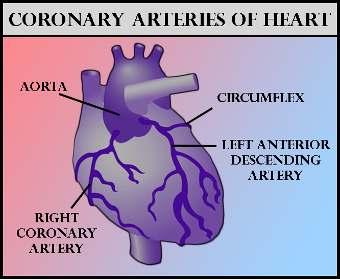
Like other muscles in the body, your heart needs blood to get oxygen and nutrients. Yourcoronary arteries supply blood to your heart. These arteries branch off from the aorta so that oxygen-rich blood is delivered to your heart as well as the rest of your body.
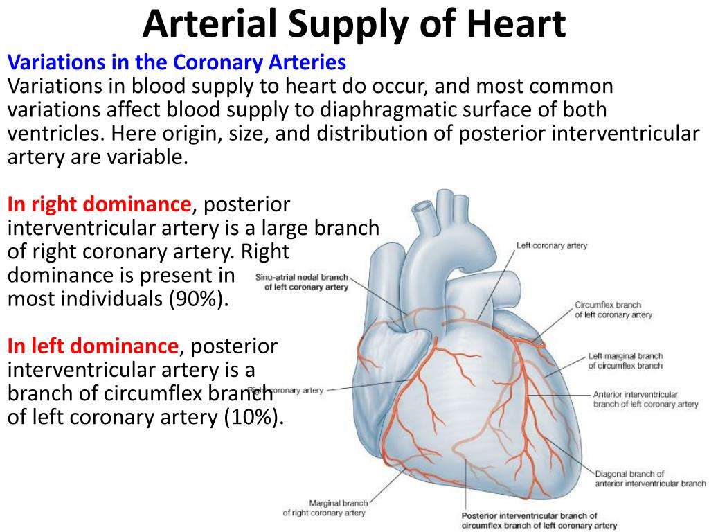
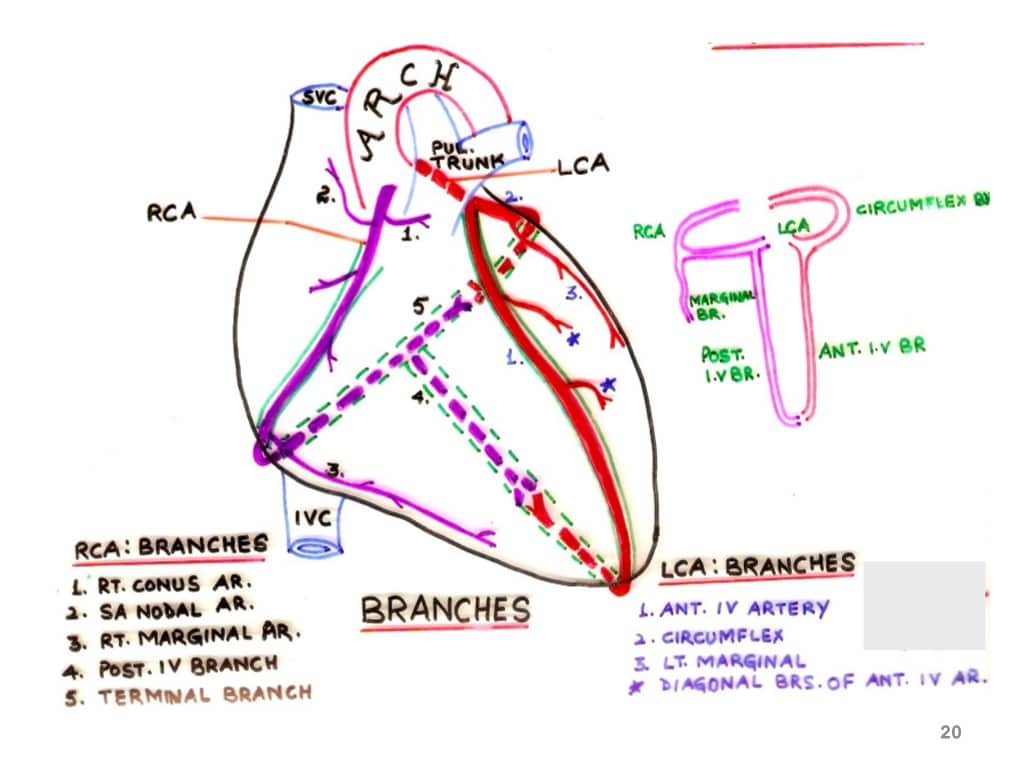
- The left coronary artery delivers blood to the left side of your heart, including your left atrium and ventricle and the septum between the ventricles.
- The circumflex artery branches off from the left coronary artery to supply blood to part of the left ventricle.
- The left anterior descending artery also branches from the left coronary artery and provides blood to parts of both the right and left ventricles.
- The right coronary artery provides blood to the right atrium and parts of both ventricles.
- The marginal arteries branch from the right coronary artery and provide blood to the surface of the right atrium.
- The posterior descending artery also branches from the right coronary artery and provides blood to the bottom of both ventricles.
Arteries supplying oxygen to the body. The coronary arteries branch off the aorta and supply the heart muscle with oxygen and nutrients. At the top of your aorta, arteries branch off to carry blood to your head and arms. Arteries branching from the middle and lower parts of your aorta supply blood to the rest of your body.
Some conditions can affect normal blood flow through these heart arteries. Examples include:
- The small cardiac vein.
Also Check: Reflux Palpitations
Blood Flow During Exercise
Blood flow within muscles fluctuates as they contract and relax. During contraction, the vasculature within the muscle is compressed, resulting in a lower arterial inflow with inflow increased upon relaxation. The opposite effect would be seen if measuring venous outflow.
This rapid increase and decrease in flow is observed over multiple contractions. If the muscle is used for an extended period, mean arterial inflow will increase as the arterioles vasodilate to provide the oxygen and nutrients required for contraction. Following the end of contractions, this increased mean flow remains to resupply the muscle tissue with required nutrients and clear inhibitory waste products, due to the loss of the inhibitory contractile phase.
Four Chambers Of The Heart And Blood Circulation

The shape of the human heart is like an upside-down pear, weighing between 7-15 ounces, and is little larger than the size of the fist. It is located between the lungs, in the middle of the chest, behind and slightly to the left of the breast bone. The heart, one of the most significant organs in the human body, is a muscular pump, which pumps blood throughout the body. It beats approximately 72 times per minute, and pumps oxygenated blood to different parts of the body.
Atria and Ventricles
There are four chambers in your heart that are left atrium, right atrium, left ventricle, and right ventricle.
- The upper chambers of your heart are atria, whereas the lower chambers are ventricles.
- Deoxygenated blood enters your heart through the right atria from where the blood moves into the right ventricle first and then to the lungs through the pulmonary artery. Your left atrium receives oxygenated blood from the lungs, which is then pumped into the left ventricle from where it moves into the aorta and then to the different parts of your body.
Valves to Ensure Unidirectional Blood Flow
Your heart has four types of valves with primary function of regulating the blood flow through the heart. Every heart diagram labeledwill clearly show these valves. These valves allow blood flow in one direction only. Different valves perform different functions.
Blood Vessels
The Circulation of Blood
Recommended Reading: Coronary Insufficiency Symptoms
