Iv Hepatic Portal System:
- A large hepatic portal vein is formed by the joining of several branches from stomach, intestine, spleen and pancreas.
- It carries blood of alimentary canal, heavily loaded with digested food stuffs, to the liver into which it breaks up into capillaries.
- The anterior abdominal vein is connected with hepatic portal vein, in the region of liver.
How Does Blood Flow Through Your Lungs
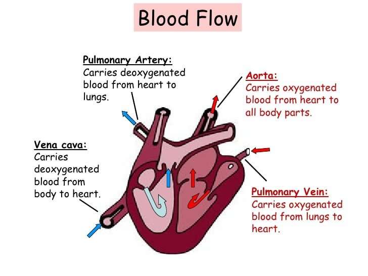
Once blood travels through the pulmonic valve, it enters your lungs. This is called the pulmonary circulation. From your pulmonic valve, blood travels to the pulmonary arteries and eventually to tiny capillary vessels in the lungs.
Here, oxygen travels from the tiny air sacs in the lungs, through the walls of the capillaries, into the blood. At the same time, carbon dioxide, a waste product of metabolism, passes from the blood into the air sacs. Carbon dioxide leaves the body when you exhale. Once the blood is oxygenated, it travels back to the left atrium through the pulmonary veins.
Summary: Distribution Of Blood Flow
The following list breaks down the blood flow throughout the body:
- Systemic circulation 84%
When blood flow needs to be redistributed to other portions of the body, the vasomotor center located in the medulla oblongata sends sympathetic stimulation to the smooth muscles in the walls of the veins, causing constrictionor in this case, venoconstriction. Less dramatic than the vasoconstriction seen in smaller arteries and arterioles, venoconstriction may be likened to a stiffening of the vessel wall. This increases pressure on the blood within the veins, speeding its return to the heart. As you will note in the image above, approximately 21 percent of the venous blood is located in venous networks within the liver, bone marrow, and integument. This volume of blood is referred to as venous reserve. Through venoconstriction, this reserve volume of blood can get back to the heart more quickly for redistribution to other parts of the circulation.
Don’t Miss: Does Dehydration Cause Increased Heart Rate
What Are The Coronary Arteries
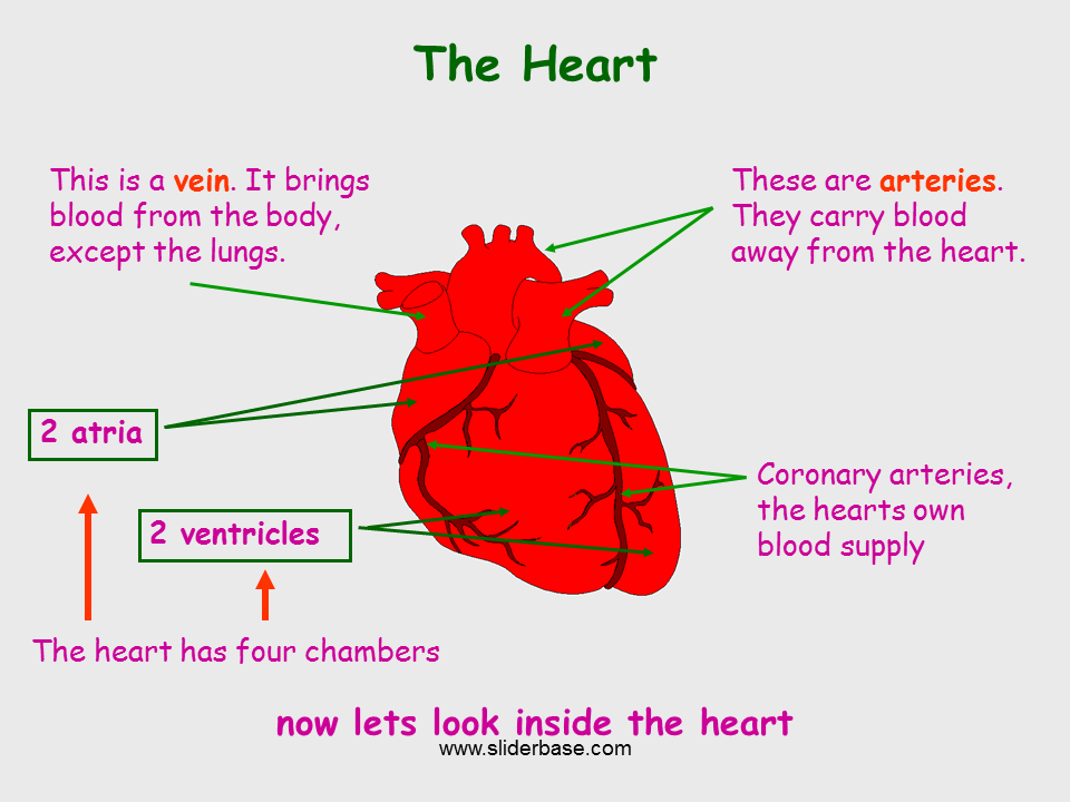
Like all organs, your heart is made of tissue that requires a supply of oxygen and nutrients. Although its chambers are full of blood, the heart receives no nourishment from this blood. The heart receives its own supply of blood from a network of arteries, called the coronary arteries.
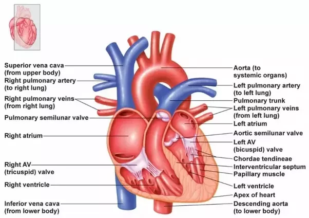
Two major coronary arteries branch off from the aorta near the point where the aorta and the left ventricle meet:
- Right coronary artery supplies the right atrium and right ventricle with blood. It branches into the posterior descending artery, which supplies the bottom portion of the left ventricle and back of the septum with blood.
- Left main coronary artery branches into the circumflex artery and the left anterior descending artery. The circumflex artery supplies blood to the left atrium, as well as the side and back of the left ventricle. The left anterior descending artery supplies the front and bottom of the left ventricle and the front of the septum with blood.
These arteries and their branches supply all parts of the heart muscle with blood.
When the coronary arteries narrow to the point that blood flow to the heart muscle is limited , a network of tiny blood vessels in the heart that aren’t usually open may enlarge and become active. This allows blood to flow around the blocked artery to the heart muscle, protecting the heart tissue from injury.
What Carries Blood To The Heart What Carries Blood Away From The Heart

Veins carry blood to the heart, arteries carry blood from the heart.
Explanation:
There are multiple veins which carry blood to the heart.
The two venae cavae carry de-oxygenated blood from the arms, head, and upper body and from the legs and lower body to the left atrium .
The four pulmonary veins carry oxygenated blood from the lungs to the right atrium .
There are two major arteries which carry blood from the heart.
The pulmonary artery carries de-oxygenated blood from the left ventricle to the lungs for oxygenation.
The aorta carries oxygenated blood from the right ventricle to the rest of the body for energy and metabolism.
Also Check: Chamber That Pushes Blood Through The Aortic Valve
Circulatory System Of Frog
- The blood vascular or circulatory system of frog is closed.
- The circulatory system consists of:
- Heart:
Exchange Of Gases Nutrients And Waste Between Blood And Tissue Occurs In The Capillaries
Capillaries are tiny vessels that branch out from arterioles to form networks around body cells. In the lungs, capillaries absorb oxygen from inhaled air into the bloodstream and release carbon dioxide for exhalation. Elsewhere in the body, oxygen and other nutrients diffuse from blood in the capillaries to the tissues they supply. The capillaries absorb carbon dioxide and other waste products from the tissues and then flow the deoxygenated blood into the veins.
Also Check: Effects Of Exercise On Cardiac Output
What Does The Circulatory System Do
The circulatory system is made up of blood vessels that carry blood away from and towards the heart. Arteries carry blood away from the heart and veins carry blood back to the heart.
The circulatory system carries oxygen, nutrients, and to cells, and removes waste products, like carbon dioxide. These roadways travel in one direction only, to keep things going where they should.
How Does The Heart Beat
The heart gets messages from the body that tell it when to pump more or less blood depending on a person’s needs. For example, when we’re sleeping, it pumps just enough to provide for the lower amounts of oxygen needed by our bodies at rest. But when we’re exercising, the heart pumps faster so that our muscles get more oxygen and can work harder.
How the heart beats is controlled by a system of electrical signals in the heart. The sinus node is a small area of tissue in the wall of the right atrium. It sends out an electrical signal to start the contracting of the heart muscle. This node is called the pacemaker of the heart because it sets the rate of the heartbeat and causes the rest of the heart to contract in its rhythm.
These electrical impulses make the atria contract first. Then the impulses travel down to the atrioventricular node, which acts as a kind of relay station. From here, the electrical signal travels through the right and left ventricles, making them contract.
One complete heartbeat is made up of two phases:
Read Also: Ibs And Heart Palpitations
What Are Three Types Of Blood Vessels
There are three kinds of blood vessels: arteries, veins, and capillaries. Each of these plays a very specific role in the circulation process. Arteries carry oxygenated blood away from the heart. Theyre tough on the outside but they contain a smooth interior layer of epithelial cells that allows blood to flow easily.
Career Connection Vascular Surgeons And Technicians
Vascular surgery is a specialty in which the physician deals primarily with diseases of the vascular portion of the cardiovascular system. This includes repair and replacement of diseased or damaged vessels, removal of plaque from vessels, minimally invasive procedures including the insertion of venous catheters, and traditional surgery. Following completion of medical school, the physician generally completes a 5-year surgical residency followed by an additional 1 to 2 years of vascular specialty training. In the United States, most vascular surgeons are members of the Society of Vascular Surgery.
Vascular technicians are specialists in imaging technologies that provide information on the health of the vascular system. They may also assist physicians in treating disorders involving the arteries and veins. This profession often overlaps with cardiovascular technology, which would also include treatments involving the heart. Although recognized by the American Medical Association, there are currently no licensing requirements for vascular technicians, and licensing is voluntary. Vascular technicians typically have an Associates degree or certificate, involving 18 months to 2 years of training. The United States Bureau of Labor projects this profession to grow by 29 percent from 2010 to 2020.
Read Also: Can Tylenol Lower Heart Rate
What Is The Vascular System
The vascular system, also called the circulatory system, is made up of the vessels that carry blood and lymph through the body. The arteries and veins carry blood throughout the body, delivering oxygen and nutrients to the body tissues and taking away tissue waste matter. The lymph vessels carry lymphatic fluid . The lymphatic system helps protect and maintain the fluid environment of the body by filtering and draining lymph away from each region of the body.
The vessels of the blood circulatory system are:
-
Arteries. Blood vessels that carry oxygenated blood away from the heart to the body.
-
Veins. Blood vessels that carry blood from the body back into the heart.
-
Capillaries. Tiny blood vessels between arteries and veins that distribute oxygen-rich blood to the body.
Blood moves through the circulatory system as a result of being pumped out by the heart. Blood leaving the heart through the arteries is saturated with oxygen. The arteries break down into smaller and smaller branches to bring oxygen and other nutrients to the cells of the body’s tissues and organs. As blood moves through the capillaries, the oxygen and other nutrients move out into the cells, and waste matter from the cells moves into the capillaries. As the blood leaves the capillaries, it moves through the veins, which become larger and larger to carry the blood back to the heart.
How Can I Help Keep My Child’s Heart Healthy

To help keep your child’s heart healthy:
- Encourage plenty of exercise.
- Help your child reach and keep a healthy weight.
- Go for regular medical checkups.
- Tell the doctor about any family history of heart problems.
Let the doctor know if your child has any chest pain, trouble breathing, or dizzy or fainting spells or if your child feels like the heart sometimes goes really fast or skips a beat.
You May Like: What Causes Bleeding Around The Heart
What Is The Cardiovascular System
The cardiovascular system, also called the circulatory system, is the organ system that transports materials to and from all the cells of the body. The materials carried by the cardiovascular system include oxygen from the lungs, nutrients from the digestive system, hormones from glands of the endocrine system, and waste materials from cells throughout the body. Transport of these and many other materials is necessary to maintain homeostasis of the body. The main components of the cardiovascular system are the heart, blood vessels, and blood. Each of these components is shown in Figure \ and introduced in the text.
What Do The Heart And Blood Vessels Do
The heart’s main function is to pump blood around the body. Blood carries nutrients and waste products and is vital to life. One of the essential nutrients found in blood is oxygen.
The right side of the heart receives blood lacking oxygen from the body. After passing through the right atrium and right ventricle this blood is pumped to the lungs. Here blood picks up oxygen and loses another gas called carbon dioxide. Once through the lungs, the blood flows back to the left atrium. It then passes into the left ventricle and is pumped into the main artery supplying the body. Oxygenated blood is then carried though blood vessels to all the body’s tissues. Here oxygen and other nutrients pass into the cells where they are used to perform the body’s essential functions.
A blood vessel’s main function is to transport blood around the body. Blood vessels also play a role in controlling your blood pressure.
Blood vessels are found throughout the body. There are five main types of blood vessels: arteries, arterioles, capillaries, venules and veins.
Arteries carry blood away from the heart to other organs. They can vary in size. The largest arteries have special elastic fibres in their walls. This helps to complement the work of the heart, by squeezing blood along when heart muscle relaxes. Arteries also respond to signals from our nervous system, either tightening or relaxing .
Read Also: Tylenol And High Blood Pressure
Disorders Of The Cardiovascular System: Edema And Varicose Veins
Despite the presence of valves and the contributions of other anatomical and physiological adaptations we will cover shortly, over the course of a day, some blood will inevitably pool, especially in the lower limbs, due to the pull of gravity. Any blood that accumulates in a vein will increase the pressure within it, which can then be reflected back into the smaller veins, venules, and eventually even the capillaries. Increased pressure will promote the flow of fluids out of the capillaries and into the interstitial fluid. The presence of excess tissue fluid around the cells leads to a condition called edema.
Most people experience a daily accumulation of tissue fluid, especially if they spend much of their work life on their feet . However, clinical edema goes beyond normal swelling and requires medical treatment. Edema has many potential causes, including hypertension and heart failure, severe protein deficiency, renal failure, and many others. In order to treat edema, which is a sign rather than a discrete disorder, the underlying cause must be diagnosed and alleviated.
Figure 7. Varicose veins are commonly found in the lower limbs.
Why Do I Feel A Pulse In My Stomach
Youre most likely just feeling your pulse in your abdominal aorta. Your aorta is the main artery that carries blood from your heart to the rest of your body. It runs from your heart, down the center of your chest, and into your abdomen. Its normal to feel blood pumping through this large artery from time to time.
Read Also: Medical Name For Enlarged Heart
Veins Carry Blood Back Toward The Heart
After the capillaries release oxygen and other substances from blood into body tissues, they feed the blood back toward the veins. First the blood enters microscopic vein branches called venules. The venules conduct the blood into the veins, which transport it back to the heart through the venae cavae. Vein walls are thinner and less elastic than artery walls. The pressure pushing blood through them is not as great. In fact, there are valves within the lumen of veins to prevent the backflow of blood.
The Constant Pumping Of The Heart Maintains Blood Pressure And Supply Throughout The Body
The blood moving through the circulatory system puts pressure on the walls of the blood vessels. Blood pressure results from the blood flow force generated by the pumping heart and the resistance of the blood vessel walls. When the heart contracts, it pumps blood out through the arteries. The blood pushes against the vessel walls and flows faster under this high pressure. When the ventricles relax, the vessel walls push back against the decreased force. Blood flow slows down under this low pressure.
Read Also: Reflux Heart Palpitations
What Are The Functions Of Blood Vessels
Blood vessels play crucial roles within the body. Their functions are:
- Transporting blood with oxygen and nutrients throughout the body.
- Removing waste products from the cells of the tissues and organs.
- Aiding in gas exchange.
- Maintaining homeostasis and health.
- Working as a network for white blood cells to allow their circulation in the body when the immune response is activated against infection or injury.
- Blood vessels near the skin play a key role in thermoregulation by increasing or decreasing blood flow.
What Is Vascular Disease

A vascular disease is a condition that affects the arteries and veins. Most often, vascular disease affects blood flow, either by blocking or weakening blood vessels, or by damaging the valves that are found in veins. Organs and other body structures may be damaged by vascular disease as a result of decreased or completely blocked blood flow.
Read Also: Can Tylenol Cause Heart Palpitations
Oxygenated Blood Flows Away From The Heart Through Arteries
The left ventricle of the heart pumps oxygenated blood into the aorta. From there, blood passes through major arteries, which branch into muscular arteries and then microscopic arterioles. The arterioles branch into the capillary networks that supply tissues with oxygen and nutrients. The walls of arteries are thicker than the walls of veins, with more smooth muscle and elastic tissue. This structure allows arteries to dilate as blood pumps through them.
Anatomy Of The Heart And Blood Vessels
Reviewed byDr Jacqueline Payne
The heart is a muscular pump that pushes blood through blood vessels around the body. The heart beats continuously, pumping the equivalent of more than 14,000 litres of blood every day through five main types of blood vessels: arteries, arterioles, capillaries, venules and veins.
Also Check: Can Tylenol Stop A Heart Attack
Structure Of Blood And Blood Vessels
Blood is carried through three different types of blood vessels in the body:
All blood vessels are specifically structured to perform their function. For example, a capillary is microscopically thin to allow gases to exchange, the arteries are tough and flexible to cope with high pressure blood flow and the veins contain valves to prevent the blood from travelling backwards when at low pressure. All vessels feature varying lumen size. The lumen is the hollow opening or the space inside the blood vessel.
| Artery | |||
|---|---|---|---|
| Carry blood away from the heart | Carry blood towards the heart | Allows diffusion of gases and nutrients from blood into the body cells | |
| Wall | Very thin, one cell thick | ||
| Lumen | Very small, only allows blood to pass through one cell at a time | ||
| Other features | Thick muscular walls to withstand blood flowing at high pressure as it leaves the heart the largest artery is the aorta | Contain valves to prevent back flow of blood | Walls are made of semi-permeable membrane to allow transport of gases and nutrients into and out of the blood |
