Brain Anatomy And Limbic System
The image on the left is a side view of the outside of the brain, showing the major lobes and the brain stem structures .
The image on the right is a side view showing the location of the limbic system inside the brain. The limbic system consists of a number of structures, including the fornix, hippocampus, cingulate gyrus, amygdala, the parahippocampal gyrus, and parts of the thalamus. The hippocampus is one of the first areas affected by Alzheimers disease. As the disease progresses, damage extends throughout the lobes.
Read Also: How To Calculate Resting Heart Rate
What Is Neural Control Of Breathing
The neural control of respiration refers to functional interactions between networks of neurons that regulate movements of the lungs, airways and chest wall and abdomen, in order to accomplish effective organismal uptake of oxygen and expulsion of carbon dioxide, airway liquids and irritants, regulation of
Free Masterclass: The Ultimate Framework To Transform Your Mind Body And Relationships
Why is life so easy for some, and so difficult for others?Have you ever wondered how some people seem to float through life effortlessly, and the things they want just flow to them as if theyre blessed by magic?What if there was a framework you could follow, that could transform your mind, body and relationships and set you up for success in any area you choose?What if there was a way to reshape your deepest beliefs about yourself, enabling you to achieve daily personal breakthroughs on a subconscious, intuitive, and automatic level?Join Mindvalley founder Vishen Lakhiani in this FREE Masterclass as he dives deep into the core personal growth practices that will insert life-changing habits into your day-to-day living so you can live the life you always wanted to live.
Recommended Reading: Does Tylenol Increase Heart Rate
Right Brain Left Brain
The cerebrum is divided into two halves: the right and left hemispheres They are joined by a bundle of fibers called the corpus callosum that transmits messages from one side to the other. Each hemisphere controls the opposite side of the body. If a stroke occurs on the right side of the brain, your left arm or leg may be weak or paralyzed.
Not all functions of the hemispheres are shared. In general, the left hemisphere controls speech, comprehension, arithmetic, and writing. The right hemisphere controls creativity, spatial ability, artistic, and musical skills. The left hemisphere is dominant in hand use and language in about 92% of people.
Components Of The Brainstem

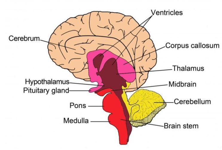
The three components of the brainstem are the medulla oblongata, midbrain, and pons.
Brainstem Anatomy: Structures of the brainstem are depicted on these diagrams, including the midbrain, pons, medulla, basilar artery, and vertebral arteries.
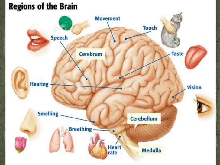
The medulla oblongata is the lower half of the brainstem continuous with the spinal cord. Its upper part is continuous with the pons. The medulla contains the cardiac, respiratory, vomiting, and vasomotor centers regulating heart rate, breathing, and blood pressure.
The midbrain is associated with vision, hearing, motor control, sleep and wake cycles, alertness, and temperature regulation.
The pons lies between the medulla oblongata and the midbrain. It contains tracts that carry signals from the cerebrum to the medulla and to the cerebellum. It also has tracts that carry sensory signals to the thalamus.
Read Also: Top Part Of Heart Not Working
What Part Of The Brain Controls Anger
Medically Reviewed By: Whitney White, MS. CMHC, NCC., LPC
We’ve known for a long time that anger and other emotions are controlled in the brain. A more recent discovery that different parts of the brain “control” different emotions.
So, what part of the brain controls anger? How do we know? What do we do with that kind of information?
Blood Supply To The Brain
Two sets of blood vessels supply blood and oxygen to the brain: the vertebral arteries and the carotid arteries.
The external carotid arteries extend up the sides of your neck, and are where you can feel your pulse when you touch the area with your fingertips. The internal carotid arteries branch into the skull and circulate blood to the front part of the brain.
The vertebral arteries follow the spinal column into the skull, where they join together at the brainstem and form the basilar artery, which supplies blood to the rear portions of the brain.
The circle of Willis, a loop of blood vessels near the bottom of the brain that connects major arteries, circulates blood from the front of the brain to the back and helps the arterial systems communicate with one another.
Also Check: Tylenol And Heart Palpitations
Where Is The Medulla Oblongata
The medulla oblongata, often simply called the medulla, is an elongated section of neural tissue that makes up part of the brainstem. The medulla is anterior to the cerebellum and is the part of the brainstem that connects to the spinal cord. It is continuous with the spinal cord, meaning there is not a clear delineation between the spinal cord and medulla but rather the spinal cord gradually transitions into the medulla.
Divisions Of The Reticular Formation
Traditionally, the nuclei are divided into three columns:
Sagittal division reveals more morphological distinctions. The raphe nuclei form a ridge in the middle of the reticular formation, and directly to its periphery, there is a division called the medial reticular formation. The medial reticular formation is large, has long ascending and descending fibers, and is surrounded by the lateral reticular formation. The lateral reticular formation is close to the motor nuclei of the cranial nerves and mostly mediates their function. The raphe nuclei is the place of synthesis of the neurotransmitter serotonin, which plays an important role in mood regulation.
The medial reticular formation and lateral reticular formation are two columns of neuronal nuclei with ill-defined boundaries that send projections through the medulla and into the mesencephalon . The nuclei can be differentiated by function, cell type, and projections of efferent or afferent nerves. The magnocellular red nucleus is involved in motor coordination, and the parvocellular nucleus regulates exhalation.
Cross Section of the Pons: A cross section of the lower part of the pons showing the pontine reticular formation labeled as #9.
Read Also: Does Tylenol Increase Heart Rate
What Part Of The Brain Controls Digestion
controlsbrainbraincontrolsdigestion
The autonomic nervous system controls the tone of the digestive tract. The brain controls drinking and feeding behavior. The brain controls muscles for eating and elimination. The digestive system sends sensory information to the brain.
One may also ask, what controls the movement of your stomach? The pyloric sphincter controls the passage of partially digested food from the stomach into the duodenum where peristalsis takes over to move this through the rest of the intestines.
Also Know, is the brain part of the digestive system?
People are most familiar with the body’s central nervous system, which is made up of the brain and spinal cord. The enteric nervous system’s network of nerves, neurons, and neurotransmitters extends along the entire digestive tract from the esophagus, through the stomach and intestines, and down to the anus.
What part of the brain controls memory?
The main parts of the brain involved with memory are the amygdala, the hippocampus, the cerebellum, and the prefrontal cortex . The amygdala is involved in fear and fear memories. The hippocampus is associated with declarative and episodic memory as well as recognition memory.
Can Female Hormones Cause Heart Palpitations
Heart palpitationsfemale hormone estrogenhearthormonecanheartandpalpitationsandStroke volume index is determined by three factors:
- Preload: The filling pressure of the heart at the end of diastole.
- Contractility: The inherent vigor of contraction of the heart muscles during systole.
- Afterload: The pressure against which the heart must work to eject blood during systole.
Recommended Reading: What Are The Early Signs Of Congestive Heart Failure
A Close Look At The Adrenal Glands
The adrenal glands are controlled in part by the brain. The hypothalamus, a small area of the brain involved in hormonal regulation, produces corticotropin-releasing hormone and vasopressin . Vasopressin and CRH trigger the pituitary gland to secrete corticotropin , which stimulates the adrenal glands to produce corticosteroids. The renin-angiotensin-aldosterone system, regulated mostly by the kidneys, causes the adrenal glands to produce more or less aldosterone is persistently high pressure in the arteries. Often no cause for high blood pressure can be identified, but sometimes it occurs as a result of an underlying read more ).
The body controls the levels of corticosteroids according to need. The levels tend to be much higher in the early morning than later in the day. When the body is stressed, due to illness or otherwise, the levels of corticosteroids increase dramatically.
Things That Can Go Wrong With The Brain

Because the brain controls just about everything, when something goes wrong with it, its often serious and can affect many different parts of the body. Inherited diseases, brain disorders associated with mental illness, and head injuries can all affect the way the brain works and upset the daily activities of the rest of the body.
Problems that can affect the brain include:
Brain tumors. A brain tumor is an abnormal tissue growth in the brain. A tumor in the brain may grow slowly and produce few symptoms until it becomes large, or it can grow and spread rapidly, causing severe and quickly worsening symptoms. Brain tumors in children can be benign or malignant. Benign tumors usually grow in one place and may be curable through surgery if theyre located in a place where they can be removed without damaging the normal tissue near the tumor. A malignant tumor is cancerous and more likely to grow rapidly and spread.
Epilepsy. This condition is made up of a wide variety of seizure disorders. Partial seizures involve specific areas of the brain, and symptoms vary depending on the location of the seizure activity. Other seizures, called generalized seizures, involve a larger portion of the brain and usually cause uncontrolled movements of the entire body and loss of consciousness when they occur. Although the specific cause is unknown in many cases, epilepsy can be related to brain injury, tumors, or infections. The tendency to develop epilepsy may be inherited in families.
You May Like: Left Ventricular Systolic Dysfunction Symptoms
Ventricles And Cerebrospinal Fluid
Deep in the brain are four open areas with passageways between them. They also open into the central spinal canal and the area beneath arachnoid layer of the meninges.
The ventricles manufacture cerebrospinal fluid, or CSF, a watery fluid that circulates in and around the ventricles and the spinal cord, and between the meninges. CSF surrounds and cushions the spinal cord and brain, washes out waste and impurities, and delivers nutrients.
Heart Rate And Heart Rate Variability In Posttraumatic Disorder
Increased heart rate is a determinant of physiological arousal seen in PTSD. Subjects who develop PTSD have been shown to have higher heart rates during the immediate aftermath of the trauma compared to traumatized individuals who do not develop PTSD. The elevated heart rate is caused by the increased noradrenergic tone, suggesting that increased noradrenergic activity immediately after the trauma may play an important role in the neurobiological processes involved in the development of PTSD. From a clinical perspective, this finding suggests that elevated heart rate immediately after the trauma is a predictor of PTSD.
J.R. Jennings, in, 2007
You May Like: Unsafe Heart Rate Resting
How The Nervous System Works
The basic functioning of the nervous system depends a lot on tiny cells called neurons. The brain has billions of them, and they have many specialized jobs. For example, sensory neurons take information from the eyes, ears, nose, tongue, and skin to the brain. Motor neurons carry messages away from the brain and back to the rest of the body.
All neurons, however, relay information to each other through a complex electrochemical process, making connections that affect the way we think, learn, move, and behave.
Intelligence, learning, and memory. At birth, the nervous system contains all the neurons you will ever have, but many of them are not connected to each other. As you grow and learn, messages travel from one neuron to another over and over, creating connections, or pathways, in the brain. Its why driving seemed to take so much concentration when you first learned but now is second nature: The pathway became established.
In young children, the brain is highly adaptable in fact, when one part of a young childs brain is injured, another part can often learn to take over some of the lost function. But as we age, the brain has to work harder to make new neural pathways, making it more difficult to master new tasks or change established behavior patterns. Thats why many scientists believe its important to keep challenging your brain to learn new things and make new connections it helps keeps the brain active over the course of a lifetime.
Show/hide Words To Know
Blood-brain barrier: a protective layer that surrounds the brain and controls what things can move into the area around the brain.
Circadian rhythm: the bodys natural clock that runs on roughly a 24 hour cycle. Many animals have a 24 hour cycle that includes sleeping, eating and doing workmore
CLSM: confocal laser scanning microscope makes high quality images of microscopic objects with extreme detailmore
Metabolism: what living things do to stay alive. This includes eating, drinking, breathing, and getting rid of wastesmore
Puberty: the change from child to adult where the body is able to reproduce.
Vertebra: any of the bones that make up the backbone.
Recommended Reading: Is Tylenol Bad For Your Heart
Disorders Of Autonomic Nervous System
People with an autonomic disorder may have trouble regulating more than one system. The common symptoms are fainting, fluctuating blood pressure, and lightheadedness.
It is a rare degenerative disorder of the autonomic nervous system. There is a general loss of autonomic functions. For example, there is reduced sweating and lacrimation, elevated blood pressure, and sexual dysfunction.
Orthostatic hypotension is the sudden drop in blood pressure on standing upright. it is a disorder in which the autonomic nervous system fails to constrict the blood vessels when a person stands up. The main complication of orthostatic hypotension is falling due to fainting.
The damage to the blood pressure sensing nerves in the neck leads to failure of the baroreflex. It causes fluctuations in the blood pressure, making it too high or too low. Symptoms of this autonomic disorder include fainting, headaches, and dizziness.
What Controls The Rate Of The Heartbeat
Heart ratecontrolledheart rate
In addition to the intrinsic heartbeat that the heart has all by itself, the autonomic nervous system is a separate part of the brain and the brain function that can either speed up or slow down your heart.
Secondly, how is heart rate controlled quizlet? At the CNS, the parasympathetic nervous system is activated and acetylcholine is released. This results in acetylcholine not being released so nothing is inhibiting the sympathetic nervous system. So Noephinephrine is released which increases the heart rate and constricts the blood vessels = increasing blood pressure.
Consequently, what part of the brain controls heart rate?
Medulla The primary role of the medulla is regulating our involuntary life sustaining functions such as breathing, swallowing and heart rate. As part of the brain stem, it also helps transfer neural messages to and from the brain and spinal cord. It is located at the junction of the spinal cord and brain.
Is a heart rate of 120 dangerous?
Well over 99 percent of the time, sinus tachycardia is perfectly normal. The increased heart rate doesnt harm the heart and doesnt require medical treatment. For example, a 10- to 15-minute brisk walk typically elevates the heart rate to 110 to 120 beats per minute.
Also Check: Unsafe Heart Rate During Exercise
Overview Of The Autonomic Nervous System
, MD, College of Medicine, Mayo Clinic
The autonomic nervous system regulates certain body processes, such as blood pressure and the rate of breathing. This system works automatically , without a persons conscious effort.
Disorders of the autonomic nervous system can affect any body part or process. Autonomic disorders may be reversible or progressive.
How Is The Heart Rate Regulated By Homeostasis

Homeostasis regulates heart rateheart rateheart rate
While heart rhythm is regulated entirely by the sinoatrial node under normal conditions, heart rate is regulated by sympathetic and parasympathetic input to the sinoatrial node. Therefore, stimulation of the accelerans nerve increases heart rate, while stimulation of the vagus nerve decreases it.
Additionally, how does homeostasis regulate blood pressure? They send impulses to the cardiovascular center to regulate blood pressure. At lower blood pressures, the degree of stretch is lower and the rate of firing is slower. When the cardiovascular center in the medulla oblongata receives this input, it triggers a reflex that maintains homeostasis.
Also Know, is heart rate an example of homeostasis?
In order for a body to work optimally, it must operate in an environment of stability called homeostasis. When the body experiences stressfor example, from exercise or extreme temperaturesit can maintain a stable blood pressure and constant body temperature in part by dialing the heart rate up or down.
What is a good resting heart rate by age?
For adults 18 and older, a normal resting heart rate is between 60 and 100 beats per minute , depending on the persons physical condition and age. For children ages 6 to 15, the normal resting heart rate is between 70 and 100 bpm, according to the AHA.
You May Like: Does Acid Reflux Cause Heart Palpitations
What Is The Medulla Oblongata And What Does It Do
For most of the 18th century, the medulla oblongata was thought to simply be an extension of the spinal cord without any distinct functions of its own. This changed in 1806, when Julien-Jean-Cesar Legallois found that he could remove the cortex and cerebellum of rabbits and they would continue to breathe. When he removed a specific section of the medulla, however, respiration stopped immediately. Legallois had found what he believed to be a “respiratory center” in the medulla, and soon after the medulla was considered to be a center of vital functions .
Over time, exactly which “vital functions” were linked to the medulla would become more clear, and the medulla would come to be recognized as a crucial area for the control of both cardiovascular and respiratory functions. The role of the medulla in cardiovascular function involves the regulation of heart rate and blood pressure to ensure that an adequate blood supply continues to circulate throughout the body at all times. To accomplish this, a nucleus in the medulla called the nucleus of the solitary tract receives information from stretch receptors in blood vessels. These receptors—called baroreceptors—can detect when the walls of blood vessels expand and contract, and thus can detect changes in blood pressure.
