Pathway Of Blood Through The Heart
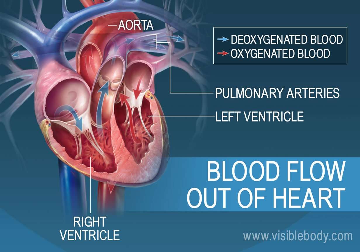
While it is convenient to describe the flow of blood through the right side of the heart and then through the left side, it is important to realize that both atria and ventricles contract at the same time. The heart works as two pumps, one on the right and one on the left, working simultaneously. Blood flows from the right atrium to the right ventricle, and then is pumped to the lungs to receive oxygen. From the lungs, the blood flows to the left atrium, then to the left ventricle. From there it is pumped to the systemic circulation.
How Does Blood Flow Through Your Lungs
Once blood travels through the pulmonic valve, it enters your lungs. This is called the pulmonary circulation. From your pulmonic valve, blood travels to the pulmonary artery to tiny capillary vessels in the lungs. Here, oxygen travels from the tiny air sacs in the lungs, through the walls of the capillaries, into the blood. At the same time, carbon dioxide, a waste product of metabolism, passes from the blood into the air sacs. Carbon dioxide leaves the body when you exhale. Once the blood is purified and oxygenated, it travels back to the left atrium through the pulmonary veins.
How Can I Protect My Heart And Pulmonary Arteries
Many of the conditions that affect the pulmonary arteries are present at birth. While you cant prevent these problems, these actions can promote better heart health:
- Eat a heart-healthy diet with plenty of fresh fruits and vegetables.
- Get at least 150 minutes of cardiovascular physical activity every week.
- Maintain a healthy weight.
Recommended Reading: Can Flonase Cause Heart Palpitations
There Are Two Types Of Circulation: Pulmonary Circulation And Systemic Circulation
Pulmonary circulation moves blood between the heart and the lungs. It transports deoxygenated blood to the lungs to absorb oxygen and release carbon dioxide. The oxygenated blood then flows back to the heart. Systemic circulation moves blood between the heart and the rest of the body. It sends oxygenated blood out to cells and returns deoxygenated blood to the heart.
The Atria Are The Hearts Entryways For Blood

The left atrium and right atrium are the two upper chambers of the heart. The left atrium receives oxygenated blood from the lungs. The right atrium receives deoxygenated blood returning from other parts of the body. Valves connect the atria to the ventricles, the lower chambers. Each atrium empties into the corresponding ventricle below.
Also Check: Afrin Heart Palpitations
The Pulmonary Loop Only Transports Blood Between The Heart And Lungs
In the pulmonary loop, deoxygenated blood exits the right ventricle of the heart and passes through the pulmonary trunk. The pulmonary trunk splits into the right and left pulmonary arteries. These arteries transport the deoxygenated blood to arterioles and capillary beds in the lungs. There, carbon dioxide is released and oxygen is absorbed. Oxygenated blood then passes from the capillary beds through venules into the pulmonary veins. The pulmonary veins transport it to the left atrium of the heart. The pulmonary arteries are the only arteries that carry deoxygenated blood, and the pulmonary veins are the only veins that carry oxygenated blood.
Function Of The Pericardium
The pericardium is important because it protects the heart from trauma, shock, stress, and even infections from the nearby lungs. It supports the heart and anchors it to the medastinum so it doesnt move within the body. The pericardium lubricates the heart and prevents it from becoming too large if blood volume is overloaded .
Despite these functions, the pericardium is still vulnerable to problems of its own. Pericarditis is the term for inflammation in the pericardium, typically due to infection. Pericarditis is often a severe disease because it can constrict and apply pressure on the heart and work against its normal function. Pericarditis comes in many types depending on which tissue layer is infected.
Don’t Miss: Does Benadryl Increase Your Heart Rate
In This Article We Explore The Structure Of The Heart How It Pumps Blood Around The Body And The Electrical System That Controls It
11+ Diagram Of The Heart Oxygenated And Deoxygenated Blood. Arteries are blood vessels that carry blood away from the heart. The walls of the ventricle contract and the blood is pushed into the pulmonary artery through the semi lunar valve which prevents blood flowing backwards into the heart. The heart pumps oxygenated blood to the body and deoxygenated blood to the lungs. Deoxygenated blood flows towards the heart.
The left side pumps oxygenated blood to the organs of the body.
From the aorta, the blood travels to various parts of the body carrying oxygen and nutrients.
This key circulatory system structure is comprised of four chambers.
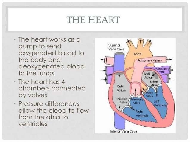
The image above shows the four chambers of the heart along with major blood vessels and valves.
The heart is a muscular organ that serves to collect deoxygenated blood from all parts of the body, carries it to the lung s to be oxygenated and release carbon the pumping of interstitial fluid from the blood into the extracellular space is an important function of the heart.
The deoxygenated blood is also called venous blood.
The heart sends deoxygenated blood to the lungs, where the blood loads up with oxygen and unloads carbon dioxide, a waste product of metabolism.
The circulatory system consists of three main types of blood vessels:
In this article, we explore the structure of the heart, how it pumps blood around the body, and the electrical system that controls it.
The Systemic Loop Goes All Over The Body
In the systemic loop, oxygenated blood is pumped from the left ventricle of the heart through the aorta, the largest artery in the body. The blood moves from the aorta through the systemic arteries, then to arterioles and capillary beds that supply body tissues. Here, oxygen and nutrients are released and carbon dioxide and other waste substances are absorbed. Deoxygenated blood then moves from the capillary beds through venules into the systemic veins. The systemic veins feed into the inferior and superior venae cavae, the largest veins in the body. The venae cavae flow deoxygenated blood to the right atrium of the heart.
Recommended Reading: Does Benadryl Lower Heart Rate
Each Heart Beat Is A Squeeze Of Two Chambers Called Ventricles
The ventricles are the two lower chambers of the heart. Blood empties into each ventricle from the atrium above, and then shoots out to where it needs to go. The right ventricle receives deoxygenated blood from the right atrium, then pumps the blood along to the lungs to get oxygen. The left ventricle receives oxygenated blood from the left atrium, then sends it on to the aorta. The aorta branches into the systemic arterial network that supplies all of the body.
The Cardiac Cycle Includes All Blood Flow Events The Heart Accomplishes In One Complete Heartbeat
The muscular wall of the heart powers contraction and dilation. Each contraction and relaxation is a heartbeat. Ventricular contractions, called systole, force blood out of the heart through the pulmonary and aortic valves. Diastole occurs when blood flows from the atria to fill the ventricles.
Read Also: Does Benadryl Lower Heart Rate
The Four Chambers Of The Heart
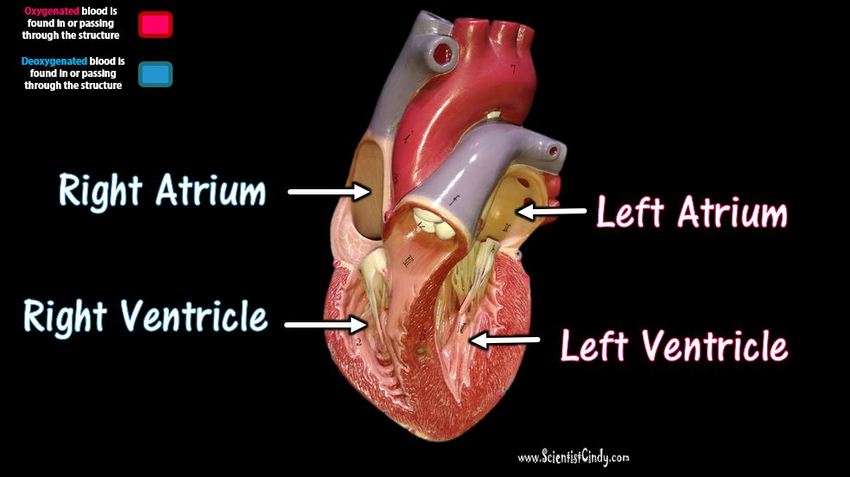
Your heart has a right and left side separated by a wall called the septum. Each side has a small collecting chamber called an atrium, which leads into a large pumping chamber called a ventricle. There are four chambers: the left atrium and right atrium , and the left ventricle and right ventricle .The right side of your heart collects blood on its return from the rest of our body. The blood entering the right side of your heart is low in oxygen. Your heart pumps the blood from the right side of your heart to your lungs so it can receive more oxygen. Once it has received oxygen, the blood returns directly to the left side of your heart, which then pumps it out again to all parts of your body through an artery called the aorta. Blood pressure refers to the amount of force the pumping blood exerts on arterial walls.
Blood Vessels Of The Heart

The blood vessels of the heart include:
- venae cavae deoxygenated blood is delivered to the right atrium by these two veins. One carries blood from the head and upper torso, while the other carries blood from the lower body
- pulmonary arteries deoxygenated blood is pumped by the right ventricle into the pulmonary arteries that link to the lungs
- pulmonary veins the pulmonary veins return oxygenated blood from the lungs to the left atrium of the heart
- aorta this is the largest artery of the body, and it runs the length of the trunk. Oxygenated blood is pumped into the aorta from the left ventricle. The aorta subdivides into various branches that deliver blood to the upper body, trunk and lower body
- coronary arteries like any other organ or tissue, the heart needs oxygen. The coronary arteries that supply the heart are connected directly to the aorta, which carries a rich supply of oxygenated blood
- coronary veins deoxygenated blood from heart muscle is ‘dumped’ by coronary veins directly into the right atrium.
Also Check: Does Acid Reflux Cause Heart Palpitations
How Your Heart Works
Your heart
The human heart is one of the hardest-working organs in the body.
On average, it beats around 75 times a minute. As the heart beats, it provides pressure so blood can flow to deliver oxygen and important nutrients to tissue all over your body through an extensive network of arteries, and it has return blood flow through a network of veins.
In fact, the heart steadily pumps an average of 2,000 gallons of blood through the body each day.
Your heart is located underneath your sternum and ribcage, and between your two lungs.
The hearts four chambers function as a double-sided pump, with an upper and continuous lower chamber on each side of the heart.
The hearts four chambers are:
- Right atrium. This chamber receives venous oxygen-depleted blood that has already circulated around through the body, not including the lungs, and pumps it into the right ventricle.
- Right ventricle. The right ventricle pumps blood from the right atrium to the pulmonary artery. The pulmonary artery sends the deoxygenated blood to the lungs, where it picks up oxygen in exchange for carbon dioxide.
- Left atrium. This chamber receives oxygenated blood from the pulmonary veins of the lungs and pumps it to the left ventricle.
- Left ventricle. With the thickest muscle mass of all the chambers, the left ventricle is the hardest pumping part of the heart, as it pumps blood that flows to the heart and rest of the body other than the lungs.
+ Diagram Of The Heart Oxygenated And Deoxygenated Blood
11+ Diagram Of The Heart Oxygenated And Deoxygenated Blood. Let’s examine the anatomy of the heart along with some diagrams that show how the heart operates. Learn vocabulary, terms and more with flashcards, games and other study tools.
An online interactive study guide to tutorials and quizzes on the anatomy and physiology of the heart, using interactive animations and diagrams. The heart pumps oxygenated blood to the body and deoxygenated blood to the lungs. Excess interstitial fluid is then.
You May Like: How To Calculate Max Hr
Blood Supply To The Myocardium
The myocardium of the heart wall is a working muscle that needs a continuous supply of oxygen and nutrients to function efficiently. For this reason, cardiac muscle has an extensive network of blood vessels to bring oxygen to the contracting cells and to remove waste products.
The right and left coronary arteries, branches of the ascending aorta, supply blood to the walls of the myocardium. After blood passes through the capillaries in the myocardium, it enters a system of cardiac veins. Most of the cardiac veins drain into the coronary sinus, which opens into the right atrium.
The Valves Are Like Doors To The Chambers Of The Heart
Four valves regulate and support the flow of blood through and out of the heart. The blood can only flow one waylike a car that must always be kept in drive. Each valve is formed by a group of folds, or cusps, that open and close as the heart contracts and dilates. There are two atrioventricular valves, located between the atrium and the ventricle on either side of the heart: The tricuspid valve on the right has three cusps, the mitral valve on the left has two. The other two valves regulate blood flow out of the heart. The aortic valve manages blood flow from the left ventricle into the aorta. The pulmonary valve manages blood flow out of the right ventricle through the pulmonary trunk into the pulmonary arteries.
Also Check: Does Tylenol Increase Heart Rate
Blood Vessels And Chambers
When you look at the orientation of the heart at the bottom of the thoracic cavity you will see that, rather than being straight up and down, the heart is at an angle, and a bit twisted . This is due in part to making room for the liver, and in part to the location of the many blood vessels that attach to the heart.
Figure 11.2 shows the blood vessels connected to the heart, but you may find the flowchart in Figure 11.3 a bit easier to understand. Don’t forget that the blood flow in the pulmonary and systemic circuits is continuous, meaning that blood from one circuit moves on immediately to the other circuit. Next, the central location of the heart means that blood going to the lungs needs to be pumped both left and right, and blood going to the body needs to be pumped both up and down. Thinking in terms of opposites will help you to remember the vessels.
Figure 11.3This flowchart illustrates the flow of blood, in terms of opposite directions, to and from both the systemic and pulmonary circuit.
Crash Cart
Flex Your Muscles
Oxygenated blood returns from the two lungs through the pulmonary veins, which attach to opposite sides of the left atrium. The rest of the trip is almost the same as on the right side: the left atrium pumps the blood through the left AV valve into the left ventricle, and the ventricle pumps the blood through the aortic semilunar valve into the aorta.
See also:
The Heart Wall Is Composed Of Three Layers
The muscular wall of the heart has three layers. The outermost layer is the epicardium . The epicardium covers the heart, wraps around the roots of the great blood vessels, and adheres the heart wall to a protective sac. The middle layer is the myocardium. This strong muscle tissue powers the hearts pumping action. The innermost layer, the endocardium, lines the interior structures of the heart.
Also Check: Acetaminophen Heart Rate
What Conditions And Disorders Affect The Pulmonary Arteries
The most common problems with the pulmonary arteries are congenital heart defects, meaning the issue is present at birth. To understand these defects you have to understand a little about the development of the heart and the circulation before you are born. Normal development of the heart and pulmonary arteries requires that the two sides of the heart shares the work equally and the way that happens is that there are two communications between the pulmonary and the systemic circulation, one atrial and one between the pulmonary artery and the aorta . Disturbed balance result in problems. These communications normally close soon after birth.
Any of these conditions can be associated with arrhythmias and cause heart failure.
Caring for Your Heart and Pulmonary Arteries
The Circulatory System Works In Tandem With The Respiratory System

The circulatory and respiratory systems work together to sustain the body with oxygen and to remove carbon dioxide. Pulmonary circulation facilitates the process of external respiration: Deoxygenated blood flows into the lungs. It absorbs oxygen from tiny air sacs and releases carbon dioxide to be exhaled. Systemic circulation facilitates internal respiration: Oxygenated blood flows into capillaries through the rest of the body. The blood diffuses oxygen into cells and absorbs carbon dioxide.
You May Like: Can Flonase Cause Heart Palpitations
The Heart Powers Both Types Of Circulation
The heart pumps oxygenated blood out of the left ventricle and into the aorta to begin systemic circulation. After the blood has supplied cells throughout the body with oxygen and nutrients, it returns deoxygenated blood to the right atrium of the heart. The deoxygenated blood shoots down from the right atrium to the right ventricle. The heart then pumps it out of the right ventricle and into the pulmonary arteries to begin pulmonary circulation. The blood moves to the lungs, exchanges carbon dioxide for oxygen, and returns to the left atrium. The oxygenated blood shoots from the left atrium to the left ventricle below, to begin systemic circulation again.
Heart Anatomy: By The Numbers
1. Superior vena cava: Receives blood from the upper body delivers blood into the right atrium.
2. Inferior vena cava: Receives blood from the lower extremities, pelvis and abdomen, and delivers blood into the right atrium.
3. Right atrium: Receives blood returning to the heart from the superior and inferior vena cava transmits blood to the right ventricle, which pumps blood to the lungs for oxygenation.
4. Tricuspid valve: Allows blood to pass from the right atrium to the right ventricle prevents blood from flowing back into the right atrium as the heart pumps .
5. Right ventricle: Receives blood from the right atrium pumps blood into the pulmonary artery.
6. Pulmonary valve: Allows blood to pass into the pulmonary arteries prevents blood from flowing back into the right ventricle.
7. Pulmonary arteries: Carry oxygen-depleted blood from the heart to the lungs.
8. Pulmonary veins: Deliver oxygen-rich blood from the lungs to the left atrium of the heart.
9. Left atrium: Receives blood returning to the heart from the pulmonary veins.
10. Mitral valve: Allows blood to flow into the left ventricle prevents blood from flowing back into the left atrium.
11. Left ventricle: Receives oxygen-rich blood from the left atrium and pumps blood into the aorta.
12. Aortic valve: Allows blood to pass from the left ventricle to the aorta prevents backflow of blood into the left ventricle.
13. Aorta: Distributes blood throughout the body from the heart.
Also Check: Ibs Heart Palpitations
