How Is Fluid Around The Lung Diagnosed
A physician will usually diagnosis pleural effusion based on interviewing the patient about symptoms and a physical examination. To confirm a diagnosis, he or she may also request an imaging test, which could be a chest X-ray, ultrasound, or computed tomography scan. To further help with diagnosis, a doctor may extract a sample of the excess fluid to be tested to determine the cause.
How Often Does Heart Surgery Cause A Pleural Effusion
Three recent medical studies shed light on this question. A study in the European Respiratory Journal analyzed patients who developed a pleural effusion after a heart surgery. About 40% of the patients developed a pleural effusion, and on average, by day seven post-op. A greater proportion of pleural effusions occurred following a coronary artery bypass graft surgery compared to heart valve replacement operations.
A similar study the American College of Chest Physicians analyzed how often patients got a pleural effusion after undergoing CABG, valve replacement, or both. Among the patients in the study, only 6.6% experienced a clinically significant pleural effusion within 30 days following the operation.
Pleural effusions were more frequent in patients with other associated cardiac conditions. Another study in the American Journal of Respiratory and Critical Care Medicine aimed to determine how often a pleural effusion developed related to the type of heart surgery. Pleural effusions in the patients undergoing only CABG surgery, or CABG plus valve surgery was higher than patients undergoing valve surgery only.
In that study, 40% of patients who had a CABG surgery developed a PE in the immediate post-op period. Most of the effusions were small, and they tended to gradually resolve themselves. Occasionally, however, the pleural effusion persisted or a new effusion developed within the first few months after surgery.
Pulmonary Edema Is A Very Serious Condition
This condition may occur after a wide range of surgical procedures and features an accumulation of fluid in the lungs which significantly affects the normal process of respiration . It amounts to a serious medical emergency.
A small amount of fluid in a person’s lungs after a surgery does not cause huge disturbances and it is much easier to deal with. On the other hand, a large amount of fluid in the lungs leads to severe shortness of breath and must be treated urgently and aggressively.
Fluid in the lungs is a potential complication of many surgeries. This is actually one of the most serious postoperative complications after certain surgical procedures. It is estimated that respiratory complications account for approximately 15% of all postoperative complications in surgeries performed under general anesthesia.
Fluid in the lungs is, for example, common after heart surgeries. In patients who have undergone a heart valve replacement surgery, pulmonary edema is one of the possible complications. In this case, it affects many patients and is, basically, an expected condition that doctors will routinely watch out for. This is why fluid in the lungs is considered more of reaction to a surgery than a complication during one.
Patients who undergo a valve replacement surgery are easily treated once the pulmonary edema occurs. It is believed that this complication occurs as a consequence of topical heart cooling which is performed with ice.
Read Also: How Much Aspirin Do You Take For A Heart Attack
Symptoms Of Fluid Around Heart And Lungs
Small amount of accumulation of fluid may remain asymptomatic however accumulation of large amount can produce range of symptoms:
Symptoms of fluid buildup around heart:
- Difficulty in breathing.
- Pain in chest, mainly in the middle of chest.
- Fever.
- Increased heart rate and pulse.
- Breathing difficulty while lying down.
- Dry cough.
- Pain is better while sitting and aggravate when lying down.
Symptoms of fluid collection around the lungs:
- Pain in chest, especially on the affected side of lungs. If fluid is collected in right side, patient experiences pain on his right side of chest. Similarly it can occur in the left side of chest.
- Difficulty while breathing.
- Pain in chest while inhaling.
- Shortness of breath.
- Loss of appetite.
- Dry cough.
Since pleural effusion or pericardial effusion is caused due to underlying disease or condition, many other symptoms of the condition are also present.
Heart Valve Repair Or Replacement Surgery

The heart is a pump made of muscle tissue. It has 4 pumping chambers: 2 upper chambers, called atria, and 2 lower chambers, called ventricles. Valves between each of the heart’s pumping chambers keep blood flowing forward through the heart.
- Tricuspid valve. Located between the right atrium and the right ventricle
- Pulmonary valve. Located between the right ventricle and the pulmonary artery
- Mitral valve. Located between the left atrium and the left ventricle
- Aortic valve. Located between the left ventricle and the aorta
When valves are damaged or diseased and do not work the way they should they may need to be repaired or replaced. Conditions that may cause heart valve dysfunction are valve stenosis and valve regurgitation .
When one valve becomes stenotic , the heart has to work harder to pump the blood through the valve. Valves can become narrow and stiff from infection and aging. If one or more valves become leaky, blood leaks backwards, which means less blood is pumped in the right direction. Based on your symptoms and the overall condition of your heart, your healthcare provider may decide that the diseased valve needs to be surgically repaired or replaced.
Read Also: Does Magnesium Help With Heart Palpitations
What Makes Yale Medicines Approach To Pleural Effusion Special
At Yale Medicine, patients receive care from a team of physicians who specialize in dealing with pleural effusions. The clinical care team includes a physician assistant and an advanced practice registered nurse who are trained in this subspecialty. What makes Yale especially unique, Dr. Puchalski adds, is our ability to perform bilateral thoracenteses. This means that a patient can have fluid build-up removed from both lung areas in a single treatment, rather than scheduling two separate procedures. Patients can do this at Yale Medicine, Dr. Puchalski explains, due to a highly-trained staff.
Another unique aspect of care at Yale Medicine is that doctors rarely ask patients to stop taking blood-thinning medication before the procedure. Many other medical centers require that patients stop blood thinners one week before the procedure, Dr. Puchalski says. However, Yale researchers conducted thorough research and found that this precaution did not affect the final outcome of the procedure. We dont make patients wait to undergo the procedure, he says.
Pulmonary Edema Vs Pneumonia
Pneumonia is another serious condition of the lungs. Unlike edema, pneumonia is caused by either a viral, fungal, or bacterial infection. As your lungs become infected, fluid builds up in the air sacs .
While both pulmonary edema and pneumonia cause a form of buildup in the lungs, the former is primarily caused by CHF. Pneumonia, on the other hand, is caused by an infection. A weakened immune system can increase your chances of getting pneumonia from a common cold or flu.
Symptoms of pneumonia may include:
- high fever with chills
- cough with mucus that continues to worsen
- chest pain and discomfort
Don’t Miss: Who Performed The First Open Heart Surgery Successful
Pleural Effusion & Heart Surgery: What Should Patients Know
You might have experienced a pleural effusion, or you may know someone who has. Pleural effusions are a common complication after heart surgery. They can be painful and cause concern. You may have questions such as, What causes a pleural effusion?, What are the symptoms?, and How is it treated?
Atelectasis And Gas Exchange After Cardiac Surgery
Senior Registrar, Department of Cardiothoracic Anesthesia.
Associate Professor, Department of Cardiothoracic Anesthesia.
Associate Professor, Department of Diagnostic Radiology.
Professor and Chair, Department of Clinical Physiology.
Anesthesiology
Arne Tenling, Thomas Hachenberg, Hans Tyden, Goran Wegenius, Goran Hedenstierna Atelectasis and Gas Exchange after Cardiac Surgery . Anesthesiology 1998 89:371378 doi:
Sometimes a high intrapulmonary shunt occurs after cardiac surgery, and impairment of lung function and oxygenation can persist for 1 week after operation. Animal studies have shown that postoperative shunt can be explained by atelectasis. In this study the authors tried to determine if atelectasis can explain shunt in patients who have had cardiac surgery.
Nine patients having coronary artery bypass graft surgery and nine patients having mitral valve surgery were examined using the multiple inert gas elimination technique before and after operation. On the first postoperative day, computed tomography scans were made at three levels of the thorax.
Large atelectasis in the dorsal part of the lungs was found on the first postoperative day after cardiac surgery. However, there was no clear correlation between atelectasis and measured shunt fraction.
Read Also: Target Heart Rate When Working Out
Treatment For Fluid Around The Heart And Lungs
Many cases of fluid buildup around the heart and lungs resolve without any specific treatment. In few other cases treating the underlying cause will improve the condition.
For example if the accumulation is caused due to tuberculosis, anti tuberculosis treatment with medications will gradually reduce excess collection of fluid.
If accumulation is in excess and the symptoms worsen, the fluid may need to be drained with a large bore needle. The procedure is performed by a trained doctor with the help of sonogram in sterile environment.
What Causes Fluid Around The Heart And Lungs
Build up of excess fluid in pericardium and pleura can result from number of causes.
Causes of fluid buildup around heart:
- Viral or bacterial infection.
- Accumulation of fluid after heart surgery or after heart attack.
- Rheumatoid arthritis or other autoimmune disease.
- Kidney and liver failure.
- Trauma or puncture wound which infiltrates the pericardium.
- Radiation therapy involving radiation of the lung tissue.
- Hypothyroidism.
- Metastatic infiltration in lung and breast cancer.
- Certain chemotherapy drugs.
- Prescription drugs such as hydralazine, isoniazide, phenytoin etc.
Causes of fluid buildup around lungs:
Abnormal fluid accumulation can occur between the layers of pleura due to several reasons.
- Infection such as tuberculosis of lungs.
- Pneumonia.
- Viral infection such as dengue fever.
- Organ failure which includes liver and kidney failure.
- Cancer of lungs.
- Metastasis occurring from breast cancer, ovarian cancer or prostate cancer.
- Autoimmune diseases such as lupus, rheumatic arthritis.
- After open heart surgery.
- Congestive cardiac failure.
Don’t Miss: Why Does Heart Rate Increase With Exercise
Fluid In Lungs After Heart Valve Surgery
By Adam Pick on December 13, 2007
Earlier today, I received an email from Stacey Ballan, a caregiver. Staceys mom recently had heart valve replacement surgery. Inside her email, there was a very interesting question that brought back memories of a minor minor heart valve surgery complication that I experienced.
Staceys email states, Adam My mother was supposed to be leaving the hospital today . However, now the doctors say they have found fluid in her lungs. Is this normal or could it mean her valve is still leaking somehow? I feel so bad for her, she was all excited about coming home. Any idea as to what may be happening?
So you know, I am not a surgeon, a cardiologist or a pulmonary specialist. That said, I can not comment on the reasons why Staceys mom is experiencing fluid in her lungs.
However, I did experience fluid in my lungs for the first week following my double heart valve replacement . It felt like a terrible cramp in my ribs that would not go away. Every time I breathed in, there would be a long, pinch of pain. Needless to say, it wasnt fun.
When I told my cardiologist about pain, Dr. Rosin told me it was most likely fluid in my lungs after bypass surgery. Dr. Rosin instructed me to use my incentive spirometer every hour for ten minutes for two days. The cardiologist assured me the pain would go away.
I hope this helps explain a little bit more about fluid in the lungs after heart bypass surgery and heart valve surgery.
Keep on tickin!
Fluid Retention After Surgery

The term ‘edema’ refers to the visible swelling that is caused by accumulation of excess fluid in the body tissues. There have been instances of edema in individuals who have undergone a surgery. This write-up will throw some light on the possible causes of fluid retention after surgery.
The term edema refers to the visible swelling that is caused by accumulation of excess fluid in the body tissues. There have been instances of edema in individuals who have undergone a surgery. This write-up will throw some light on the possible causes of fluid retention after surgery.
Accumulation of fluid in the interstitial spaces of bodys organs could be caused due to a wide range of reasons. It could be a symptom of serious medical conditions such as heart failure, kidney problems, thyroid problems, diabetes, metabolic disorders or chronic venous insufficiency. Lymphatic obstruction, which is a medical condition that is characterized by the inability of the lymphatic vessels to drain lymph fluid from the body tissues due to a blockage in lymphatic vessels, could also cause lymphedema. Lymphedema could be caused due to injuries or infections. It could even develop as a post-surgery complication owing to the damage caused to lymphatic vessels during surgery.
You May Like: What To Take For Heart Palpitations
To Help You Balance Your Fluids After Heart Surgery Your Doctor May Ask You To:
- Avoid adding salt when cooking.
- Avoid processed foods.
- Read food labels for sodium content.
- Eat a low-salt diet .
- Walk daily as advised.
- Take a medication to remove excess fluid if needed.
Other symptoms of increased fluid include decrease in energy level and dizziness.
It is important to notify your doctor or nurse as soon as possible to prevent any problems related to excess fluid.
For more information: See your Guide to Cardiac Surgery binder. If you have any questions or concerns, please call the Heart, Vascular & Thoracic Institute Post Discharge Phone Line at the number you were provided or your doctors office.
This information is about care at Cleveland Clinic and may include instructions specific to Cleveland Clinic Heart, Vascular & Thoracic Institute patients only. Please consult your physician for information pertaining to your care.
Anesthesia And Mechanical Ventilation
Morphine and scopolamine were given intramuscularly 1 h before anesthesia. Anesthesia and muscle relaxation was induced with 5 – 10 g/kg fentanyl, 1.5 – 2.5 mg/kg thiopental, and 0.1 mg/kg pancuronium. After tracheal intubation, mechanical ventilation was started with 50% oxygen in nitrogen. Tidal volume and respiratory frequency were adjusted to maintain a normal arterial carbon dioxide tension. No positive end-expiratory pressure was used during the study.
Anesthesia was maintained with small doses of fentanyl and inhaled isoflurane . The lungs were not kept inflated during cold cardioplegic arrest. By the time the aortic cross clamp was removed, ventilation was resumed with 100% oxygen and one half the tidal volume of that before bypass. For the MVR patients, air was also expelled from the heart and central circulation by several deep breaths .
In the intensive care unit, mechanical ventilation was maintained as described before with an inspired oxygen fraction of 0.35 – 0.5, tidal volume of 8 – 10 ml/kg, and a respiratory frequency of 10 – 14 breaths/min. Patients were tracheally extubated when they were awake and hemodynamically stable, had a carbon dioxide tension < 5.5 kPa while spontaneously breathing, an oxygen tension > 10 kPa , an inspired oxygen fraction < 0.45, and a body temperature > 37 C. All patients received 1 – 5 l/min supplementary oxygen until 20 min before the final measurements were made.
Also Check: What Is Heart Valve Disease
How Is Fluid Around The Lung Treated
The best way is to treat the cause of the effusion. If the cause is pneumonia, a doctor will likely prescribe antibiotics to treat the infection, which may also cause the fluid to go away. If fluid build-up has been caused by congestive heart failure, a physician will likely prescribe diuretics, such as Lasix, for treatment.
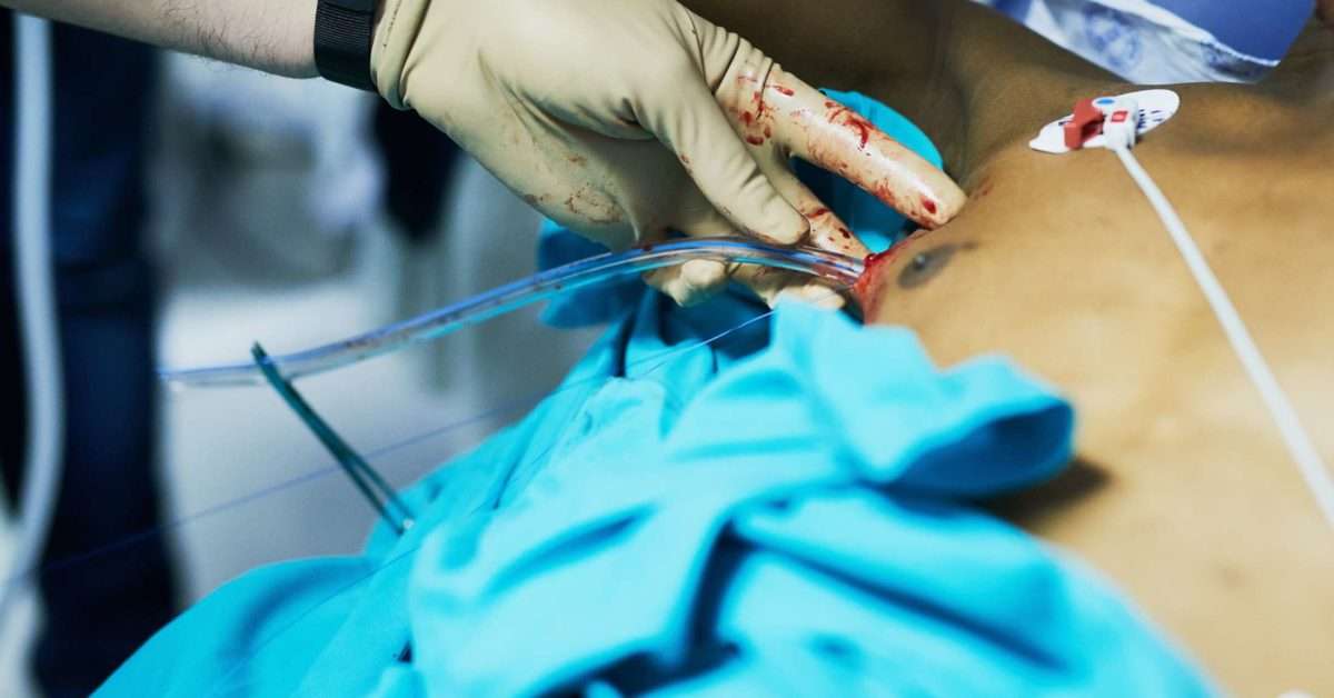
For large pleural effusions, or for those with an unknown cause, the fluid will need to be drained through a procedure called thoracentesis. This involves inserting a needle in the space between the lung and the chest wall and draining the liquid. In these cases, a doctor may also send a sample of fluid to be tested for other causes, such as lung cancer, for example. Some patients may require a pleural drain that is inserted through the skin so that the buildup of fluid can be drained repeatedly without the need for repeated thoracentesis.
What Is Pleural Effusion
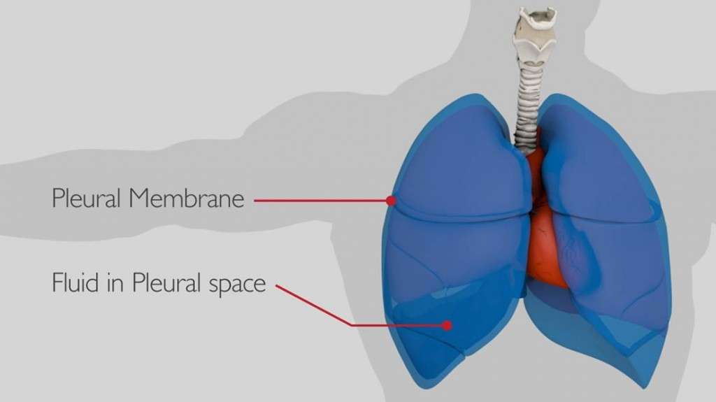
Pleural effusion, also called water on the lung, happens when fluid builds up in the space between your lungs and chest cavity.
Thin membranes, called pleura, cover the outside of the lungs and the inside of the chest cavity. Theres always a small amount of liquid within this lining to help lubricate the lungs as they expand within the chest during breathing. However, if too much fluid builds up, for example, because of a medical condition, problems can arise. Doctors call this pleural effusion.
Various conditions can lead to pleural effusion, but congestive heart failure is the
- chest pain
- weight loss
The doctor may drain the fluid or carry out pleurodesis if youre likely to need repeated drainage. This involves insering a shunt that redirects the fluid away from the chest.
They may prescribe antibiotics if you have or are susceptible to infection. Steroids or other anti-inflammatory medications may reduce pain and inflammation. They will also discuss other treatment options for cancer.
People who are undergoing treatment for cancer may also have compromised immune systems, making them more prone to infections or other complications.
The treatment and outcome will depend on the cause of the pleural effusion.
You May Like: How Does A Heart Attack Occur
Pleural Effusion Post Coronary Artery Bypass Surgery: Associations And Complications
John D. L. Brookes1^, Michael Williams1,2^, Manish Mathew1, Tristan Yan1,2,3,4,5^, Paul Bannon1,2,5,6
1Department of Cardiothoracic Surgery, 3Professor of Cardiovascular and Thoracic Surgery, Macquarie University 4Clinical Professor of Surgery, Faculty of Medicine, The University of Sydney 6Bosch Professor of Surgery, Faculty of Medicine, The University of Sydney , Australia
Contributions: Conception and design: All authors Administrative support: None Provision of study materials or patients: None Collection and assembly of data: JDL Brookes, M Williams Data analysis and interpretation: JDL Brookes, M Williams Manuscript writing: All authors Final approval of manuscript: All authors.
^ORCID: John D. L. Brookes, 0000-0002-8568-6130 Michael Williams, 0000-0002-7002-9728 Tristan Yan, 0000-0002-7473-1522.
Correspondence to:
Background: One of the most frequent complications of coronary artery bypass grafting is pleural effusion. Limited previous studies have found post-CABG pleural effusion to be associated with increased length-of-stay and greater morbidity post-CABG. Despite this the associations of this common complication are poorly described. This study sought to identify modifiable risk factors for effusion post-CABG.
Methods: A retrospective cohort study of prospectively collected data assessed patients who underwent CABG over two-years. Data was collected for risk factors and sequelae related to pleural effusion requiring drainage.
doi: 10.21037/jtd-20-2082
