How Veins Work Theory
How veins work
Once the blood has travelled through the arteries into the capillaries , the oxygen and nutrients are transferred to the tissues and are replaced with waste products, including carbon dioxide, water and urea. The venous blood at the ankle just after going through the capillaries has just enough pressure left in it to push it back to the heart. Therefore if we spend all our time lying on our backs, the veins would have very little work to do .
Therefore the veins work by movement of the legs, pumping the blood up and out of the veins.
The veins themselves do not do any pumping. They are passive blood vessels which are very elastic and can distend when full with blood, but can be easily compressed allowing blood to be pushed out of them.
It is thanks to the pumping of blood upwards during active contraction of the muscles, followed by the closure of the valve stopping the blood falling back down when the muscles relax that make sure that blood only flows one-way in veins and continues upwards towards the heart.
What Does The Heart Do
The heart is a pump, usually beating about 60 to 100 times per minute. With each heartbeat, the heart sends blood throughout our bodies, carrying oxygen to every cell. After delivering the oxygen, the blood returns to the heart. The heart then sends the blood to the lungs to pick up more oxygen. This cycle repeats over and over again.
Blood And Blood Vessels
Blood, the heart and the vessels that carry blood around the body together make up the cardiovascular system. They are vital for carrying nutrients, oxygen and waste around the body.
Blood is made of cells and plasma. There are 3 main types of blood cells red cells, white cells and platelets. All are made in the marrow found in many bones.
Red blood cells deliver oxygen from the lungs to the rest of the body, and carry waste products to be released by the lungs or the kidneys. Red blood cells contain haemoglobin, which is the protein that binds and releases oxygen.
White blood cells are part of the immune system. They detect and fight infections or foreign molecules that enter the body.
Platelets are small cells that help the blood clot.
Plasma is the clearish fluid that carries the cells. It also carries the nutrients from our diet such as sugars, fats, proteins, vitamins and minerals.
As well as carrying cells, nutrients, oxygen and waste, blood also helps to regulate body temperature.
Recommended Reading: Acid Reflux Cause Palpitations
Which Conditions Affect The Venous System
Many conditions can affect your venous system. Some of the most common ones include:
- Deep vein thrombosis . A blood clot forms in a deep vein, usually in your leg. This clot can potentially travel to your lungs, causing pulmonary embolism.
- Superficial thrombophlebitis. An inflamed superficial vein, usually in your leg, develops a blood clot. While the clot can occasionally travel to a deep vein, causing DVT, thrombophlebitis is generally less serious than DVT.
- Varicose veins. Superficial veins near the surface of the skin visibly swell. This happens when one-way valves break down or vein walls weaken, allowing blood to flow backward.
- Chronic venous insufficiency. Blood collects in the superficial and deep veins of your legs due to improper functioning of one-way valves. While similar to varicose veins, chronic venous insufficiency usually causes more symptoms, including coarse skin texture and ulcers in some cases.
While the symptoms of a venous condition can vary widely, some common ones include:
- inflammation or swelling
- veins that feel warm to the touch
- a burning or itching sensation
These symptoms are especially common in your legs. If you notice any of these and they dont improve after a few days, make an appointment with your doctor.
They can perform a venography. In this procedure, your doctor injects contrast die into your veins to produce an X-ray image of a particular area.
What Does The Circulatory System Do

The circulatory system is made up of blood vessels that carry blood away from and towards the heart. Arteries carry blood away from the heart and veins carry blood back to the heart.
The circulatory system carries oxygen, nutrients, and to cells, and removes waste products, like carbon dioxide. These roadways travel in one direction only, to keep things going where they should.
Recommended Reading: Can Acid Reflux Cause Heart Palpitations
Understanding The Circulation System : By What Mechanism Does Blood Return To The Heart
Have you ever wondered or even thought about this question? Most of our lives we are in an upright position, either standing or sitting.
Blood is a liquid and, like all liquids on the surface of the earth, blood conforms to the law of gravity. Throw a glass of water into the air and it reliably falls to the earth. A physical force acts upon the water pulls it to the earth.
Dealing with gravity is simply a fact of life here on earth. So when you assume an upright position in the morning the force of gravity immediately begins to act on the liquids in your body and draw them back to the earth.
Veins Carry Blood Back Toward The Heart
After the capillaries release oxygen and other substances from blood into body tissues, they feed the blood back toward the veins. First the blood enters microscopic vein branches called venules. The venules conduct the blood into the veins, which transport it back to the heart through the venae cavae. Vein walls are thinner and less elastic than artery walls. The pressure pushing blood through them is not as great. In fact, there are valves within the lumen of veins to prevent the backflow of blood.
Don’t Miss: Can Too Much Vitamin D Cause Heart Palpitations
What Are The Coronary Arteries
Like all organs, your heart is made of tissue that requires a supply of oxygen and nutrients. Although its chambers are full of blood, the heart receives no nourishment from this blood. The heart receives its own supply of blood from a network of arteries, called the coronary arteries.
Two major coronary arteries branch off from the aorta near the point where the aorta and the left ventricle meet:
- Right coronary artery supplies the right atrium and right ventricle with blood. It branches into the posterior descending artery, which supplies the bottom portion of the left ventricle and back of the septum with blood.
- Left main coronary artery branches into the circumflex artery and the left anterior descending artery. The circumflex artery supplies blood to the left atrium, as well as the side and back of the left ventricle. The left anterior descending artery supplies the front and bottom of the left ventricle and the front of the septum with blood.
These arteries and their branches supply all parts of the heart muscle with blood.
When the coronary arteries narrow to the point that blood flow to the heart muscle is limited , a network of tiny blood vessels in the heart that aren’t usually open may enlarge and become active. This allows blood to flow around the blocked artery to the heart muscle, protecting the heart tissue from injury.
The Constant Pumping Of The Heart Maintains Blood Pressure And Supply Throughout The Body
The blood moving through the circulatory system puts pressure on the walls of the blood vessels. Blood pressure results from the blood flow force generated by the pumping heart and the resistance of the blood vessel walls. When the heart contracts, it pumps blood out through the arteries. The blood pushes against the vessel walls and flows faster under this high pressure. When the ventricles relax, the vessel walls push back against the decreased force. Blood flow slows down under this low pressure.
Recommended Reading: How Much Blood Does The Heart Pump
The Three Major Types Of Blood Vessels: Arteries Veins And Capillaries
Blood vessels flow blood throughout the body. Arteries transport blood away from the heart. Veins return blood back toward the heart. Capillaries surround body cells and tissues to deliver and absorb oxygen, nutrients, and other substances. The capillaries also connect the branches of arteries and to the branches of veins. The walls of most blood vessels have three distinct layers: the tunica externa, the tunica media, and the tunica intima. These layers surround the lumen, the hollow interior through which blood flows.
Blood Vessels: Circulating The Blood
|
Blood travels from the heart in arteries, which branch into smaller and smaller vessels, eventually becoming arterioles. Arterioles connect with even smaller blood vessels called capillaries. Through the thin walls of the capillaries, oxygen and nutrients pass from blood into tissues, and waste products pass from tissues into blood. From the capillaries, blood passes into venules, then into veins to return to the heart. Arteries and arterioles have relatively thick muscular walls because blood pressure in them is high and because they must adjust their diameter to maintain blood pressure and to control blood flow. Veins and venules have much thinner, less muscular walls than arteries and arterioles, largely because the pressure in veins and venules is much lower. Veins may dilate to accommodate increased blood volume. |
If a blood vessel breaks, tears, or is cut, blood leaks out, causing bleeding. Blood may flow out of the body, as external bleeding, or it may flow into the spaces around organs or directly into organs, as internal bleeding.
Read Also: Does Acid Reflux Cause Heart Palpitations
The Blood Supply To The Heart
Like any other muscle, the heart muscle needs a good blood supply. The coronary arteries take blood to the heart muscle. These are the first arteries to branch off the large artery which takes blood to the body from the left ventricle.
- The right coronary artery mainly supplies the muscle of the right ventricle.
- The left coronary artery quickly splits into two and supplies the rest of the heart muscle.
- The main coronary arteries divide into many smaller branches to supply all the heart muscle.
The Venous Return Curve

If right atrial pressure were changed in steps over the entire range of possible atrial pressures and venous return were measured at each point, plotting the data set would yield a complete venous return curve, which is presented in . As mentioned earlier, such measurements would have to be made during total blockade of the autonomic nervous system so that circulatory reflexes would be normal. Notice that, at the normal right atrial pressure value , venous return is 100%, which is 5 L/min in man. Venous return falls progressively as right atrial pressure increases, until right atrial pressure reaches 7 mm Hg, the normal value for mean systemic pressure. At that point, venous return is 0 because the pressure gradient for venous return is 0. As right atrial pressure falls below 0, the venous return curve increases at a progressively declining rate until flow reaches a plateau at approximately 4 mm Hg. As discussed above, the reason for the curvilinear nature in this portion of the relationship, termed the transition zone, is the progressive increase in vascular resistance due to the collapse of increasing numbers of veins as right atrial pressure becomes more negative.
The complete venous return curve over the range of right atrial pressure from 8 to 8 mm Hg. Venous return values are for humans.
Recommended Reading: Can Reflux Cause Heart Palpitations
Mechanisms To Return Blood
The return of blood to the heart is assisted by the action of the skeletal-muscle pump and by the thoracic pump action of breathing during respiration. As muscles move, they squeeze the veins that run through them. Veins contain a series of one-way valves. As the vein is squeezed, it pushes blood through the valves, which then close to prevent backflow. Standing or sitting for prolonged periods can cause low venous return from venous pooling. In venous pooling, the smooth muscles surrounding the veins become slack and the veins fill with the majority of the blood in the body, keeping blood away from the brain, which can cause unconsciousness.
Venous valve: Venous valves prevent back flow and ensure that blood flows in one direction.
Although most veins take blood back to the heart, portal veins carry blood between capillary beds. For example, the hepatic portal vein takes blood from the capillary beds in the digestive tract and transports it to the capillary beds in the liver. The blood is then drained in the gastrointestinal tract and spleen, where it is taken up by the hepatic veins and blood is taken back into the heart. Since this is an important function in mammals, damage to the hepatic portal vein can be dangerous. Blood clotting in the hepatic portal vein can cause portal hypertension, which results in a decrease of blood fluid to the liver.
Where Are The Heart And Blood Vessels Found
The heart is a fist-sized organ which lies within the chest behind the breastbone . The heart sits on the main muscle of breathing , which is found beneath the lungs. The heart is considered to have two ‘sides’ – the right side and the left side.
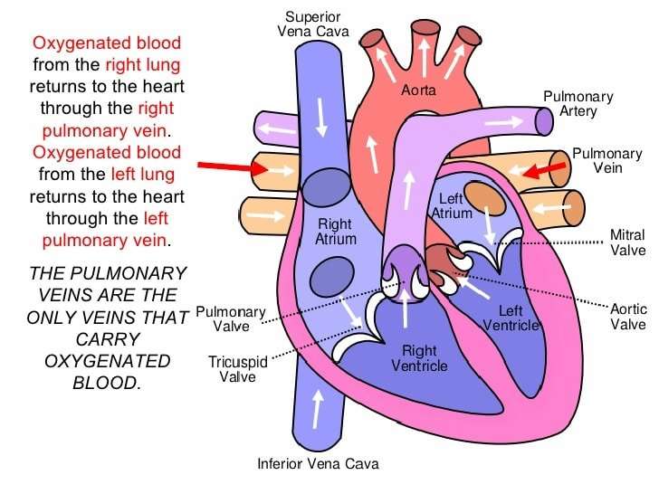
The heart has four chambers – an atrium and a ventricle on each side. The atria are both supplied by large blood vessels that bring blood to the heart . Atria have special valves that open into the ventricles. The ventricles also have valves but, in this case, they open into blood vessels. The walls of the heart chambers are made mainly of special heart muscle. The different sections of the heart have to squeeze in the correct order for the heart to pump blood efficiently with each heartbeat.
Recommended Reading: How To Calculate Resting Heart Rate
There Are Three Main Types Of Blood Vessels
Arteries
The arteries carry oxygen and nutrients away from your heart, to your body’s tissues.The veins take oxygen-poor blood back to the heart.
- Arteries begin with the aorta, the large artery leaving the heart.
- They carry oxygen-rich blood away from the heart to all of the body’s tissues.
- They branch several times, becoming smaller and smaller as they carry blood further from the heart.
Capillaries
- Capillaries are small, thin blood vessels that connect the arteries and the veins.
- Their thin walls allow oxygen, nutrients, carbon dioxide and waste products to pass to and from the tissue cells.
Veins
- These are blood vessels that take oxygen-poor blood back to the heart.
- Veins become larger and larger as they get closer to the heart.
- The superior vena cava is the large vein that brings blood from the head and arms to the heart, and the inferior vena cava brings blood from the abdomen and legs into the heart.
This vast system of blood vessels – arteries, veins, and capillaries – is over 60,000 miles long. That’s long enough to go around the world more than twice!
Blood flows continuously through your body’s blood vessels. Your heart is the pump that makes it all possible.
If The Saphenous Vein Is Removed How Is The Body Able To Circulate Blood
Small veins give deoxigenated blood to the main saphenous vein. After that, deoxygenated blood goes through the saphenous to the heart and lungs to get fresh oxygen and circulate it through arteries. If you remove the saphenous, how does this process work? How is the body able to circulate blood without it?
A healthy saphenous vein assists in returning blood from the lower extremities to the heart, but this function is dependent on the presence of a healthy system of check valves throughout the vein.In the presence of venous insufficiency, where the valves are not working, blood runs in the wrong direction from the body down to the lower leg and actually overloads the other veins that are trying to return blood to the heart.Under normal circumstances, 70-80% of the blood returning to the heart comes through the deep veins in the muscles of the leg. In the circumstances of saphenous vein reflux or varicose veins, these deep veins have to work extra hard to return the blood to the heart. After removal or ablation of a diseased saphenous vein, the blood will flow through the deep system in a normal manner. In most instances, the deep system has more than enough excess capacity to handle the blood that would normally go through the saphenous vein.
The saphenous vein is not a major vein and, in fact, provides only a tiny amount of venous return. It is a superficial vein and all of the superficial veins in the lower leg only account for 10% of venous return.
Don’t Miss: Does Acid Reflux Cause Heart Palpitations
Anatomy Of Veins And Arteries
The walls of veins and arteries are both made up of three layers:
- Outer. Tunica adventitia is the outer layer of a blood vessel, including arteries and veins. Its mostly composed of collagen and elastic fibers. These fibers enable the veins and arteries to stretch a limited amount. They stretch enough to be flexible while maintaining stability under the pressure of blood flow.
- Middle. The middle layer of the walls of arteries and veins is called the tunica media. Its made of smooth muscle and elastic fibers. This layer is thicker in arteries and thinner in veins.
- Inner. The inner layer of the blood vessel wall is called tunica intima. This layer is made of elastic fiber and collagen. Its consistency varies based on the type of blood vessel.
Unlike arteries, veins contain valves. Veins need valves to keep the blood flowing toward the heart. Theses valves are particularly important in the legs and arms. They fight gravity to prevent the backflow of blood.
Arteries dont need valves because the pressure from the heart keeps the blood flowing through them in one direction.
