Blood Flow Positive And Negative Effects
A healthy heart normally beats anywhere from 60 to 70 times per minute when you’re at rest. This rate can be higher or lower depending on your health and physical fitness; athletes generally have a lower resting heart rate, for example.
Your heart rate rises with physical activity, as your muscles consume oxygen while they work. The heart works harder to bring oxygenated blood where it is needed.
Disrupted or irregular heartbeats can affect blood flow through the heart. This can happen in multiple ways:
- Electrical impulses that regulate your heartbeat are impacted, causing an arrythmia, or irregular heartbeat. Atrial fibrillation is a common form of this.
- Conduction disorders, or heart blocks, affect the cardiac conduction system, which regulates how electrical impulses move through the heart. The type of blockan atrioventricular block or bundle branch blockdepends on where it occurs in the conduction system.
- Damaged or diseased valves can become ineffective or leak blood in the wrong direction.
- A blocked blood vessel, which can happen gradually or suddenly, can disrupt blood flow, such as during a heart attack.
If you experience an irregular heartbeat or cardiac symptoms like chest pain and shortness of breath, seek medical help immediately.
Pulmonary Circulation And Systemic Circulation
Now that you know of the basic process of how does blood flow through the heart, let us learn about the two main circulatory paths of your cardiovascular system: pulmonary and systemic circulation.
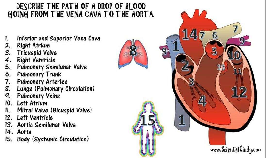
Pulmonary circulation refers to the bloods movement from your heart towards the lungs for its oxygenation and then moving back to your heart. Deoxygenated blood enters your right atrium via the inferior and superior vena cavae. Your blood gets pumped to your right ventricle via the tricuspid valve. The blood then moves to the pulmonary artery via your pulmonary valve. The big pulmonary artery divides into the left and right pulmonary arteries that travel to each of your two lungs.
On reaching your lungs, the blood moves via the capillary beds resting on the numerous alveoli. Respiration takes place and carbon dioxide is removed from the blood and is replaced with oxygen. Alveoli are basically the tiny air sacs or pouches in your lungs that provide the surface needed for the exchange of gases. The fresh blood containing oxygen leaves your lungs via the pulmonary veins that return it to your left atrium. On reaching the left atrium, the pulmonary circulation becomes complete.
Detailed explanation of blood flow through the heart:
The Left Side Of The Heart
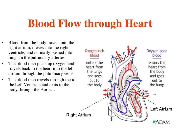
Oxygen-rich blood from the lungs passes through the pulmonary veins . It enters the left atrium and is pumped into the left ventricle. From the left ventricle, the blood is pumped to the rest of the body through the aorta.
Like all of the organs, the heart needs blood rich with oxygen. This oxygen is supplied through the coronary arteries as its pumped out of the hearts left ventricle.
The coronary arteries are located on the hearts surface at the beginning of the aorta. The coronary arteries carry oxygen-rich blood to all parts of the heart.
Also Check: How Does Exercise Affect Heart Rate
What Heart Rate Is Too High
Maximum heart rate and Target Heart Rate
Going beyond your maximum heart rate is not healthy for you. Your maximum heart rate depends on your age. This is how you can calculate it:
- Subtracting your age from the number 220 will give you your maximum heart rate. Suppose your age is 35 years, your maximum heart rate is 185 beats per minute. If your heart rate exceeds 185 beats per minute during exercise, it is dangerous for you.
- Your target heart rate zone is the range of heart rate that you should aim for if you want to become physically fit. It is calculated as 60 to 80 percent of your maximum heart rate.
- Your target heart rate helps you to know if you are exercising at the right intensity.
- It is always better to consult your doctor before starting any vigorous exercise. This is especially important if you have diabetes, heart disease, or you are a smoker. Your doctor might advise you to lower your target heart rate by 50 percent or more.
Right Side Of The Heart

- Blood enters the heart through two large veins, the inferior and superior vena cava, emptying oxygen-poor blood from the body into the right atrium of the heart.
- As the atrium contracts, blood flows from your right atrium into your right ventricle through the open tricuspid valve.
- When the ventricle is full, the tricuspid valve shuts. This prevents blood from flowing backward into the atria while the ventricle contracts.
- As the ventricle contracts, blood leaves the heart through the pulmonic valve, into the pulmonary artery and to the lungs, where it is oxygenated and then returns to the left atrium through the pulmonary veins.
You May Like: When To Go To The Hospital For Rapid Heart Rate
The Journey Of A Red Blood Cell
6 October 2017
Red Blood Cells , are cellular components of blood. There are millions of them within the human body and their sole purpose is to carry oxygen from the lungs to tissues throughout the body, as well as carrying carbon dioxide to the lungs so it can be exhaled. The blood cell is characterised by a red colour due to the presence of hemoglobin, which is a protein that helps bind oxygen to the cell.;
The red blood cell goes through a complex journey through the body, going from a deoxygenated blood cell to an oxygenated blood cell, and entering the heart twice. Below, weve laid out the journey of a red blood cell in the human body:
Supplying Oxygen To The Hearts Muscle
Like other muscles in the body, your heart needs blood to get oxygen and nutrients. Yourcoronary arteries supply blood to your heart. These arteries branch off from the aorta so that oxygen-rich blood is delivered to your heart as well as the rest of your body.
- The left coronary artery delivers blood to the left side of your heart, including your left atrium and ventricle and the septum between the ventricles.
- The circumflex artery branches off from the left coronary artery to supply blood to part of the left ventricle.
- The left anterior descending artery also branches from the left coronary artery and provides blood to parts of both the right and left ventricles.
- The right coronary artery provides blood to the right atrium and parts of both ventricles.
- The marginal arteries branch from the right coronary artery and provide blood to the surface of the right atrium.
- The posterior descending artery also branches from the right coronary artery and provides blood to the bottom of both ventricles.
Arteries supplying oxygen to the body. The coronary arteries branch off the aorta and supply the heart muscle with oxygen and nutrients. At the top of your aorta, arteries branch off to carry blood to your head and arms. Arteries branching from the middle and lower parts of your aorta supply blood to the rest of your body.
Some conditions can affect normal blood flow through these heart arteries. Examples include:
- The small cardiac vein.
Recommended Reading: Which Of The Following Body System Responses Correlates With Systolic Heart Failure (hf)
How Does Blood Flow Through The Heart
The cardiovascular or circulatory system is an important organ system of your body as it makes the blood flow through the heart, so it can transport oxygen, hormones, carbon dioxide, blood cells and nutrients to and from your body cells for nourishing the body, helping it fight diseases, stabilizing pH and temperature and maintaining homeostasis.For all these activities to take place, it is important the heart blood flow remains smooth and constant. Let us find out more about it.
Heart Diagram Parts Location And Size
Location and size of the heart
Normal heart anatomy and physiology
Normal heart anatomy and physiology need the atria and ventricles to work sequentially, contracting and relaxing to pump blood out of the heart and then to let the chambers refill. When blood leaves each chamber of the heart, it passes through a valve that is designed to prevent backflow of blood. There are four heart valves within the heart:
- Mitral valve between the left atrium and left ventricle
- Tricuspid valve between the right atrium and right ventricle
- Aortic valve between the left ventricle and aorta
- Pulmonic valve between the right ventricle and pulmonary artery
How the heart valves work
Recommended Reading: Why Does Left Arm Go Numb During Heart Attack
Pathway Of Blood Through The Heart
In this educational lesson, we learn about the blood flow order through the human heart in 14 easy steps, from the superior and inferior vena cava to the atria and ventricles. Come also learn with us the hearts anatomy, including where deoxygenated and oxygenated blood flow, in the superior vena cava, inferior vena cava, atrium, ventricle, aorta, pulmonary arteries, pulmonary veins, and coronary arteries.
Research For Your Health
The NHLBI is part of the U.S. Department of Health and Human Services National Institutes of Health the Nations biomedical research agency that makes important scientific discoveries to improve health and save lives. We are committed to advancing science and translating discoveries into clinical practice to promote the prevention and treatment of heart, lung, blood, and sleep disorders, including heart conditions. Learn about current and future NHLBI efforts to improve health through research and scientific discovery.
Read Also: Does Your Heart Rate Increase When Pregnant
What Happens During The Ablation Procedure
After you become drowsy, the doctor injects medication to numb the catheter insertion sites. The doctor inserts several catheters into large veins in both sides of your groin and possibly your neck. The catheters are advanced to the heart.
Two of the catheters are guided into the left atrium through a small hole made with a needle and placed in the atrial septum .
A transducer is inserted through one of the catheters. This is used for intracardiac ultrasound. The ultrasound lets the doctor see the structures of the heart and correctly position the catheters.
A catheter in the left atrium is used to find the abnormal impulses coming from the pulmonary veins. Another catheter is used to deliver the radiofrequency energy outside and around the pulmonary veins.
How Blood Flows Through The Heart And Lungs

The heart is a complex organ, using four chambers, four valves, and multiple blood vessels to provide blood to the body. Blood flow itself is equally complex, involving a cyclic series of steps that move blood trough the heart and to the lungs to be oxygenated, deliver it throughout the body, then bring blood back to the heart to re-start the process.
This is the key function of the cardiovascular system: consuming, transporting, and using oxygen throughout physical activity . Disruptions in blood flow through the heart and lungs can have serious effects.
You May Like: How To Calculate Your Resting Heart Rate
How The Heart Works
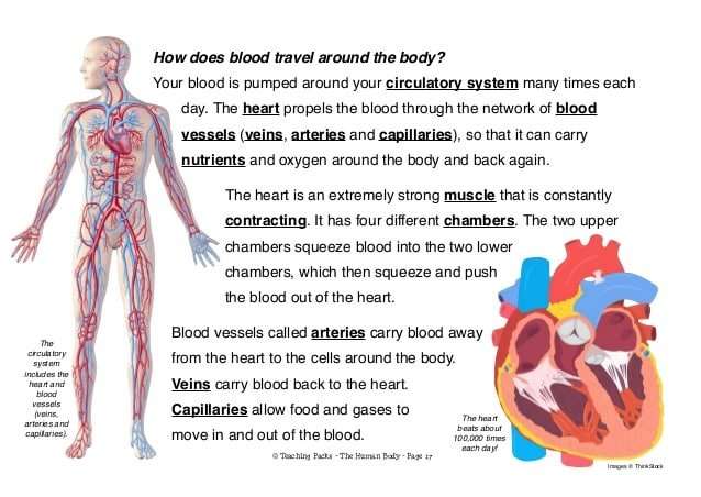
The heart is an organ, about the size of a fist. It is made of muscle and pumps blood through the body. Blood is carried through the body in blood vessels, or tubes, called arteries and veins. The process of moving blood through the body is called circulation. Together, the heart and vessels make up the cardiovascular system.
What Does The Circulatory System Do
The circulatory system is made up of blood vessels that carry blood away from and towards the heart. Arteries carry blood away from the heart and veins carry blood back to the heart.
The circulatory system carries oxygen, nutrients, and to cells, and removes waste products, like carbon dioxide. These roadways travel in one direction only, to keep things going where they should.
Also Check: What Are Symptoms Of Heart Attack
How Effective Is The Ablation Procedure In Treating Atrial Fibrillation
Cleveland Clinic has extensive experience with atrial fibrillation ablation procedures.We carefully track our patients to be certain our data are accurate.
Success rate for single ablation procedure
The success rate for a single pulmonary vein ablation procedure depends on several factors. The highest cure rate is achieved in patients with paroxysmal atrial fibrillation in whom atrial fibrillation stops on its own within 1 to 3 days. Between 75 and 80 percent of these patients whose atrial fibrillation is not related to any other heart disease are completely cured with one pulmonary vein ablation procedure.
A single ablation procedure is less likely to cure patients who have had atrial fibrillation constantly for months or years and in patients who have extensive scarring in the atrium because of other heart disease. Nonetheless, patients with long-standing atrial fibrillation can be cured with a success rate of 50 to 70 percent, depending on their underlying heart disease and other factors. These patients are more likely to require more than 1 ablation procedure.
Success rate for repeat ablation procedure
Long-term treatment goal
The long-term goal of the pulmonary vein ablation procedure is to eliminate the need for medications to prevent atrial fibrillation. Most patients can stop taking an anticoagulant a few months after the procedure because their risk of stroke is lessened.
Experience is Important
What Can I Expect After The Procedure
Discussing the procedure results: After the procedure, the doctor will discuss the results of the procedure with you and your family.
Overnight hospital stay: You will be admitted to the hospital and stay overnight for observation. Most patients go home from the hospital the next morning.
Your heart rate and rhythm will be constantly monitored during your recovery. This is done using telemetry. A a small box is connected by wires to your chest with sticky electrode patches. The box sends the information about your heart rhythm to several monitors in the nursing unit.
The doctor will remove the catheters and put pressure on the insertion site to prevent bleeding. No stitches are needed. Bandages are placed on the insertion sites to reduce the risk of bleeding and bruising.
You will stay in bed for 6 to 8 hours after the procedure. Keep your legs still during this time to prevent bleeding.
Don’t Miss: Why Does Your Heart Rate Go Up When You Exercise
What Are The Parts Of The Circulatory System
Two pathways come from the heart:
- The pulmonary circulation is a short loop from the heart to the lungs and back again.
- The systemic circulation carries blood from the heart to all the other parts of the body and back again.
In pulmonary circulation:
- The pulmonary artery is a big artery that comes from the heart. It splits into two main branches, and brings blood from the heart to the lungs. At the lungs, the blood picks up oxygen and drops off carbon dioxide. The blood then returns to the heart through the pulmonary veins.
In systemic circulation:
How The Heart Beats
How does the heart beat? Before each beat, your heart fills with blood. Then its muscle contracts to squirt the blood along. When the heart contracts, it squeezes try squeezing your hand into a fist. That’s sort of like what your heart does so it can squirt out the blood. Your heart does this all day and all night, all the time. The heart is one hard worker!
Page 1
Also Check: Does Fever Increase Heart Rate
Who Should Have Pulmonary Vein Ablation
Pulmonary vein ablation may be the best treatment option for patients who:
- Still have symptoms of atrial fibrillation, even after treatment with medications
- Cannot tolerate antiarrhythmic drugs, or have had complications from these drugs
Research has shown that atrial fibrillation usually begins in the pulmonary veins or at the point where they attach to the left atrium. There are four major pulmonary veins. All may trigger atrial fibrillation.
Patients who have treatments for atrial fibrillation often ask about Left Atrial Appendage Closure.
S Of Blood Flow Through The Heart

In summary from the video, in 14 steps, blood flows through the heart in the following order: 1) body > 2) inferior/superior vena cava > 3) right atrium > 4) tricuspid valve > 5) right ventricle > 6) pulmonary arteries >7) lungs > 8) pulmonary veins > 9) left atrium > 10) mitral or bicuspid valve > 11) left ventricle > 12) aortic valve > 13) aorta > 14) body.
Also Check: Does Sodium Increase Heart Rate
Heart Contraction And Blood Flow
Almost everyone has heard the real or recorded sound of a heartbeat. When the heart beats, it makes a lub-DUB sound. Between the time you hear lub and DUB, blood is pumped through the heart and circulatory system.
A heartbeat may seem like a simple event repeated over and over. A heartbeat actually is a complicated series of very precise and coordinated events that take place inside and around the heart.
Each side of the heart uses an inlet valve to help move blood between the atrium and ventricle.
The tricuspid valve does this between the right atrium and ventricle. The mitral valve does this between the left atrium and ventricle. The lub is the sound of the mitral and tricuspid valves closing.
Each of the hearts ventricles has an outlet valve. The right ventricle uses the pulmonary valve to help move blood into the pulmonary arteries. The left ventricle uses the aortic valve to do the same for the aorta. The DUB is the sound of the aortic and pulmonary valves closing.
Each heartbeat has two basic parts: diastole and atrial and ventricular systole . During diastole, the atria and ventricles of the heart relax and begin to fill with blood.
At the end of diastole, the hearts atria contract and pump blood into the ventricles. The atria then begin to relax. Next, the hearts ventricles contract and pump blood out of the heart.
Blood Flow During Exercise
Blood flow within muscles fluctuates as they contract and relax. During contraction, the vasculature within the muscle is compressed, resulting in a lower arterial inflow with inflow increased upon relaxation. The opposite effect would be seen if measuring venous outflow.
This rapid increase and decrease in flow is observed over multiple contractions. If the muscle is used for an extended period, mean arterial inflow will increase as the arterioles vasodilate to provide the oxygen and nutrients required for contraction. Following the end of contractions, this increased mean flow remains to resupply the muscle tissue with required nutrients and clear inhibitory waste products, due to the loss of the inhibitory contractile phase.
Recommended Reading: How Do You Calculate Max Heart Rate
