Heart Anatomy And Circulation
The heart is located in the thoracic cavity in a central compartment of the cavity known as the mediastinum. It is situated between the left and right lungs in the chest cavity. The heart is divided into upper and lower chambers called atria and ventricles . These chambers function to collect blood returning to the heart from circulation and to pump blood out of the heart. The heart is a major structure of the cardiovascular system as it serves to drive blood to all the cells of the body. Blood is circulated along a pulmonary circuit and a systemic circuit. The pulmonary circuit involves the transport of blood between the heart and lungs, while the systemic circuit involves blood circulation between the heart and the rest of the body.
A Structure That Conveys Blood From The Heart To The Lungs For Oxygenation
-
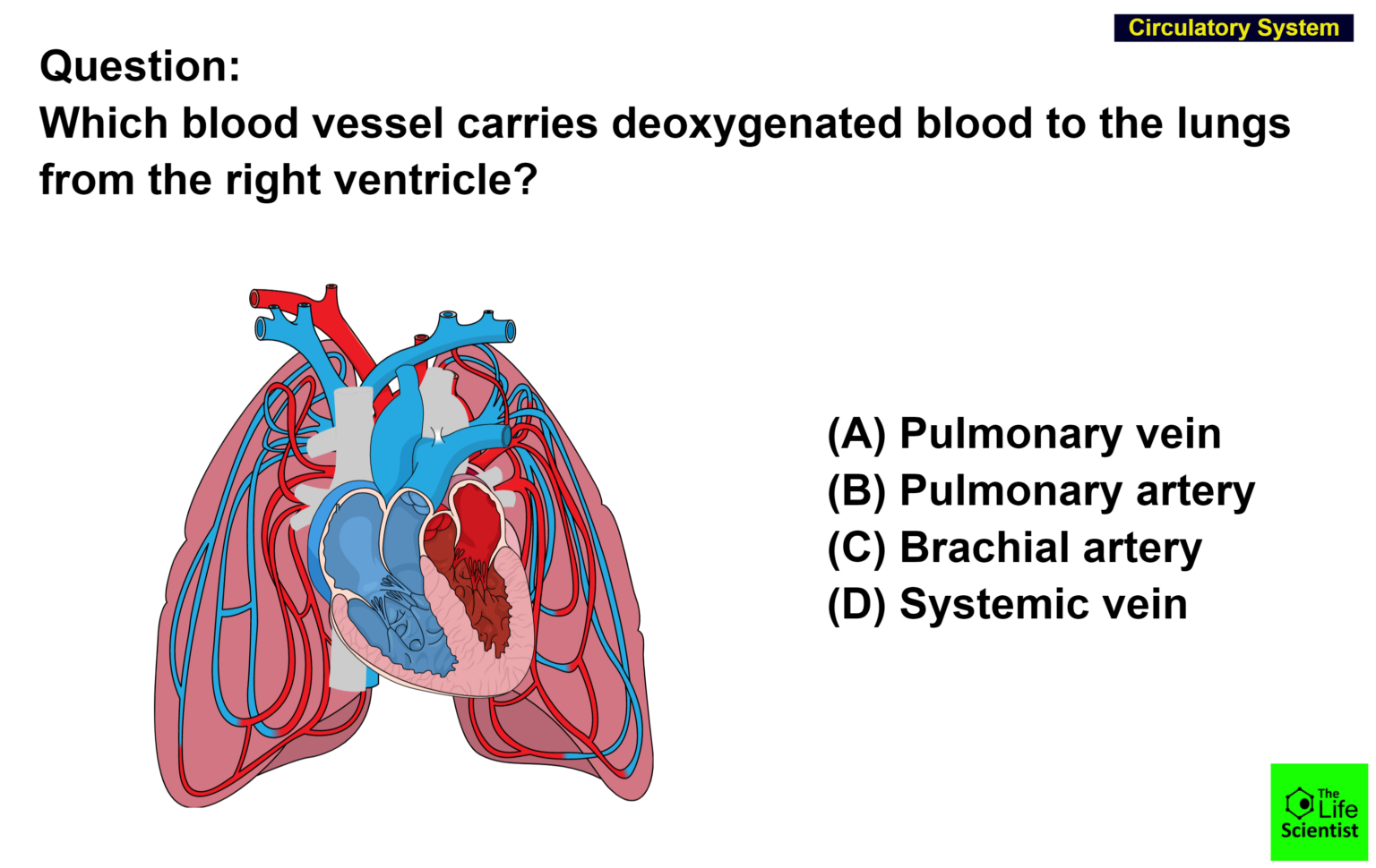
pulmonary artery
Explanation:
A blood vessel that carries blood from the heart to the lungs, where the blood picks up oxygen and then returns to the heart.
- Réponse publiée par: tayisThis statement is; false.
- Réponse publiée par: stacy05
The Social Credit System ;is a national reputation system being developed by the Chinese government.The program initiated regional trials in 2009, before launching a national pilot with eight credit scoring firms in 2014. In 2018, these efforts were centralized under the People’s Bank of China with participation from the eight firms.By 2020, it is intended to standardize the assessment of citizens’ and businesses’ economic and social reputation, or ‘Social Credit’
The Pulmonary Loop Only Transports Blood Between The Heart And Lungs
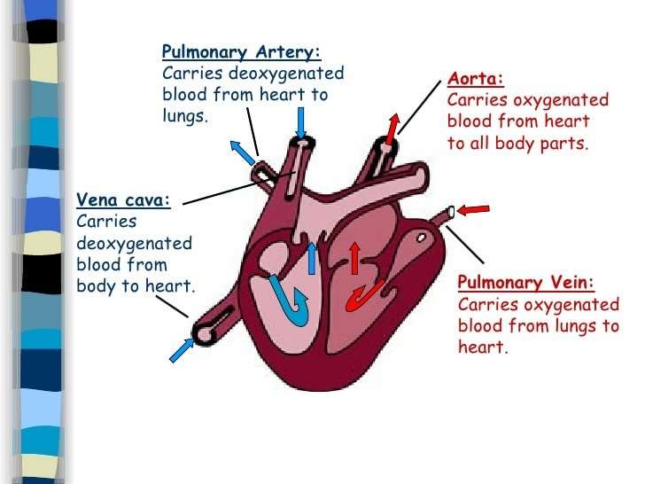
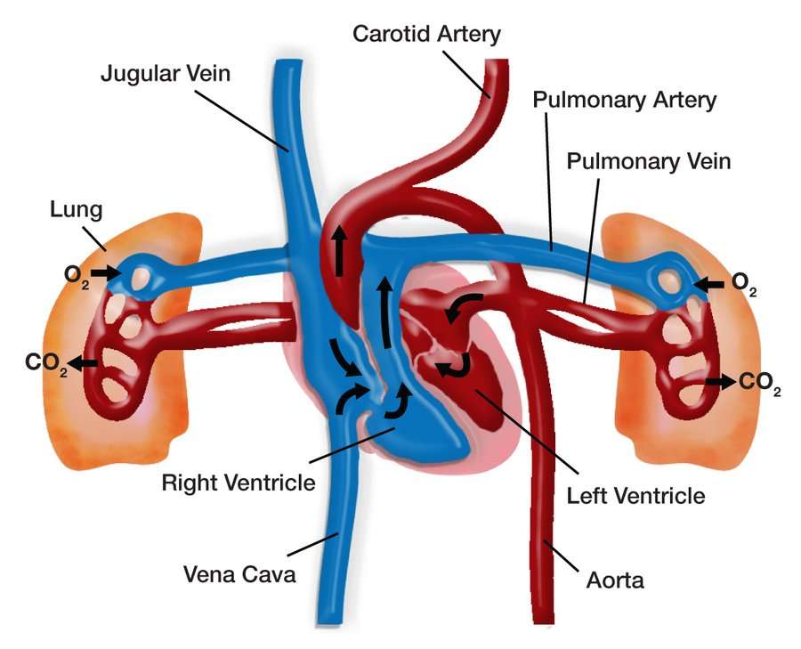
In the pulmonary loop, deoxygenated blood exits the right ventricle of the heart and passes through the pulmonary trunk. The pulmonary trunk splits into the right and left pulmonary arteries. These arteries transport the deoxygenated blood to arterioles and capillary beds in the lungs. There, carbon dioxide is released and oxygen is absorbed. Oxygenated blood then passes from the capillary beds through venules into the pulmonary veins. The pulmonary veins transport it to the left atrium of the heart. The pulmonary arteries are the only arteries that carry deoxygenated blood, and the pulmonary veins are the only veins that carry oxygenated blood.
Recommended Reading: Acid Reflux Heart Fluttering
Anatomy Of Veins And Arteries
The walls of veins and arteries are both made up of three layers:
- Outer. Tunica adventitia is the outer layer of a blood vessel, including arteries and veins. Its mostly composed of collagen and elastic fibers. These fibers enable the veins and arteries to stretch a limited amount. They stretch enough to be flexible while maintaining stability under the pressure of blood flow.
- Middle. The middle layer of the walls of arteries and veins is called the tunica media. Its made of smooth muscle and elastic fibers. This layer is thicker in arteries and thinner in veins.
- Inner. The inner layer of the blood vessel wall is called tunica intima. This layer is made of elastic fiber and collagen. Its consistency varies based on the type of blood vessel.
Unlike arteries, veins contain valves. Veins need valves to keep the blood flowing toward the heart. Theses valves are particularly important in the legs and arms. They fight gravity to prevent the backflow of blood.
Arteries dont need valves because the pressure from the heart keeps the blood flowing through them in one direction.
There Are Two Types Of Circulation: Pulmonary Circulation And Systemic Circulation

Pulmonary circulation moves blood between the heart and the lungs. It transports deoxygenated blood to the lungs to absorb oxygen and release carbon dioxide. The oxygenated blood then flows back to the heart. Systemic circulation moves blood between the heart and the rest of the body. It sends oxygenated blood out to cells and returns deoxygenated blood to the heart.
Don’t Miss: How Do You Calculate Max Heart Rate
Structure Of The Heart
The heart has four chambers . There is a wall between the two atria and another wall between the two ventricles. Arteries and veins go into and out of the heart. Arteries carry blood away from the heart and veins carry blood to the heart. The flow of blood through the vessels and chambers of the heart is controlled by valves.
What Are The Parts Of The Heart
The heart has four chambers two on top and two on bottom:
- The two bottom chambers are the right ventricle and the left ventricle. These pump blood out of the heart. A wall called the interventricular septum is between the two ventricles.
- The two top chambers are the right atrium and the left atrium. They receive the blood entering the heart. A wall called the interatrial septum is between the atria.
The atria are separated from the ventricles by the atrioventricular valves:
- The tricuspid valve separates the right atrium from the right ventricle.
- The mitral valve separates the left atrium from the left ventricle.
Two valves also separate the ventricles from the large blood vessels that carry blood leaving the heart:
- The pulmonic valve is between the right ventricle and the pulmonary artery, which carries blood to the lungs.
- The aortic valve is between the left ventricle and the aorta, which carries blood to the body.
Also Check: How To Calculate Resting Heart Rate
Heart Anatomy: By The Numbers
1. Superior vena cava: Receives blood from the upper body; delivers blood into the right atrium.
2. Inferior vena cava: Receives blood from the lower extremities, pelvis and abdomen, and delivers blood into the right atrium.
3. Right atrium: Receives blood returning to the heart from the superior and inferior vena cava; transmits blood to the right ventricle, which pumps blood to the lungs for oxygenation.
4. Tricuspid valve: Allows blood to pass from the right atrium to the right ventricle; prevents blood from flowing back into the right atrium as the heart pumps .
5. Right ventricle: Receives blood from the right atrium; pumps blood into the pulmonary artery.
6. Pulmonary valve: Allows blood to pass into the pulmonary arteries; prevents blood from flowing back into the right ventricle.
7. Pulmonary arteries: Carry oxygen-depleted blood from the heart to the lungs.
8. Pulmonary veins: Deliver oxygen-rich blood from the lungs to the left atrium of the heart.
9. Left atrium: Receives blood returning to the heart from the pulmonary veins.
10. Mitral valve: Allows blood to flow into the left ventricle; prevents blood from flowing back into the left atrium.
11. Left ventricle: Receives oxygen-rich blood from the left atrium and pumps blood into the aorta.
12. Aortic valve: Allows blood to pass from the left ventricle to the aorta; prevents backflow of blood into the left ventricle.
13. Aorta: Distributes blood throughout the body from the heart.
The Interior Of The Heart
Below is a picture of the inside of a normal, healthy, human heart.
The illustration shows a cross-section of a healthy heart and its inside structures. The blue arrow shows the direction in which low-oxygen blood flows from the body to the lungs. The red arrow shows the direction in which oxygen-rich blood flows from the lungs to the rest of the body.
You May Like: Acid Reflux Cause Palpitations
The Exterior Of The Heart:
Below is a picture of the outside of a normal, healthy, human heart.
The illustration shows the front surface of the heart, including the coronary arteries and major blood vessels.
The heart is the muscle in the lower half of the picture. The heart has four chambers. The right and left atria are shown in purple. The right and left ventricles are shown in red.
Connected to the heart are some of the main blood vessels arteries and veins that make up the blood circulatory system.
The ventricle on the right side of the heart pumps blood from the heart to the lungs.
When you breathe air in, oxygen passes from the lungs through blood vessels where its added to the blood. Carbon dioxide, a waste product, is passed from the blood through blood vessels to the lungs and is removed from the body when you breathe air out.
The atrium on the left side of the heart receives oxygen-rich blood from the lungs.
The pumping action of the left ventricle sends this oxygen-rich blood through the aorta to the rest of the body.
What Conditions And Disorders Affect The Pulmonary Arteries
The most common problems with the pulmonary arteries are congenital heart defects, meaning the issue is present at birth. To understand these defects you have to understand a little about the development of the heart and the circulation before you are born. Normal development of the heart and pulmonary arteries requires that the two sides of the heart shares the work equally and the way that happens is that there are two communications between the pulmonary and the systemic circulation, one atrial and one between the pulmonary artery and the aorta . Disturbed balance result in problems. These communications normally close soon after birth.
Any of these conditions can be associated with arrhythmias and cause heart failure.
Caring for Your Heart and Pulmonary Arteries
Don’t Miss: Can Too Much Vitamin D Cause Heart Palpitations
The Constant Pumping Of The Heart Maintains Blood Pressure And Supply Throughout The Body
The blood moving through the circulatory system puts pressure on the walls of the blood vessels. Blood pressure results from the blood flow force generated by the pumping heart and the resistance of the blood vessel walls. When the heart contracts, it pumps blood out through the arteries. The blood pushes against the vessel walls and flows faster under this high pressure. When the ventricles relax, the vessel walls push back against the decreased force. Blood flow slows down under this low pressure.
You May Like: Can Too Much Vitamin D Cause Heart Palpitations
How Does Blood Flow Through Your Lungs

Once blood travels through the pulmonic valve, it enters your lungs. This is called the pulmonary circulation. From your pulmonic valve, blood travels to the pulmonary arteries and eventually to tiny capillary vessels in the lungs.
Here, oxygen travels from the tiny air sacs in the lungs, through the walls of the capillaries, into the blood. At the same time, carbon dioxide, a waste product of metabolism, passes from the blood into the air sacs. Carbon dioxide leaves the body when you exhale. Once the blood is oxygenated, it travels back to the left atrium through the pulmonary veins.
Read Also: Does Tylenol Increase Heart Rate
What Is Pulmonary Atresia
Pulmonary atresia is a birth defect of the pulmonary valve, which is the valve that controls blood flow from the right ventricle to the main pulmonary artery . Pulmonary atresia is when this valve didnt form at all, and no blood can go from the right ventricle of the heart out to the lungs. Because a baby with pulmonary atresia may need surgery or other procedures soon after birth, this birth defect is considered a critical congenital heart defect . Congenital means present at birth.
In a baby without a congenital heart defect, the right side of the heart pumps oxygen-poor blood from the heart to the lungs through the pulmonary artery. The blood that comes back from the lungs is oxygen-rich and can then be pumped to the rest of the body. In babies with pulmonary atresia, the pulmonary valve that usually controls the blood flowing through the pulmonary artery is not formed, so blood is unable to get directly from the right ventricle to the lungs.
Disorders Of The Cardiovascular System: Edema And Varicose Veins
Despite the presence of valves and the contributions of other anatomical and physiological adaptations we will cover shortly, over the course of a day, some blood will inevitably pool, especially in the lower limbs, due to the pull of gravity. Any blood that accumulates in a vein will increase the pressure within it, which can then be reflected back into the smaller veins, venules, and eventually even the capillaries. Increased pressure will promote the flow of fluids out of the capillaries and into the interstitial fluid. The presence of excess tissue fluid around the cells leads to a condition called edema.
Most people experience a daily accumulation of tissue fluid, especially if they spend much of their work life on their feet . However, clinical edema goes beyond normal swelling and requires medical treatment. Edema has many potential causes, including hypertension and heart failure, severe protein deficiency, renal failure, and many others. In order to treat edema, which is a sign rather than a discrete disorder, the underlying cause must be diagnosed and alleviated.
Figure 7. Varicose veins are commonly found in the lower limbs.
You May Like: Does Tylenol Raise Blood Pressure And Heart Rate
The Venous Return Curve
If right atrial pressure were changed in steps over the entire range of possible atrial pressures and venous return were measured at each point, plotting the data set would yield a complete venous return curve, which is presented in . As mentioned earlier, such measurements would have to be made during total blockade of the autonomic nervous system so that circulatory reflexes would be normal. Notice that, at the normal right atrial pressure value , venous return is 100%, which is 5 L/min in man. Venous return falls progressively as right atrial pressure increases, until right atrial pressure reaches 7 mm Hg, the normal value for mean systemic pressure. At that point, venous return is 0 because the pressure gradient for venous return is 0. As right atrial pressure falls below 0, the venous return curve increases at a progressively declining rate until flow reaches a plateau at approximately 4 mm Hg. As discussed above, the reason for the curvilinear nature in this portion of the relationship, termed the transition zone, is the progressive increase in vascular resistance due to the collapse of increasing numbers of veins as right atrial pressure becomes more negative.
The complete venous return curve over the range of right atrial pressure from 8 to 8 mm Hg. Venous return values are for humans.
You May Like: How Does Anemia Cause Heart Failure
Research For Your Health
The NHLBI is part of the U.S. Department of Health and Human Services National Institutes of Health the Nations biomedical research agency that makes important scientific discoveries to improve health and save lives. We are committed to advancing science and translating discoveries into clinical practice to promote the prevention and treatment of heart, lung, blood, and sleep disorders, including heart conditions. Learn about current and future NHLBI efforts to improve health through research and scientific discovery.
You May Like: How Does Anemia Cause Heart Failure
What Circuit Connects The Heart And Lungs
pulmonary circulation
Systemic circulation
Also, how does blood circulate through the heart? Blood enters the heart through two large veins, the inferior and superior vena cava, emptying oxygen-poor blood from the body into the right atrium. As the ventricle contracts, blood leaves the heart through the pulmonic valve, into the pulmonary artery and to the lungs where it is oxygenated.
In this manner, what is the systemic circuit of the heart?
Systemic CircuitSystemic circulation carries oxygenated blood from the left ventricle, through the arteries, to the capillaries in the tissues of the body. From the tissue capillaries, the deoxygenated blood returns through a system of veins to the right atrium of the heart.
How does blood flow through the pulmonary circuit?
Pulmonary circulation is the movement of blood from the heart to the lungs for oxygenation, then back to the heart again. The blood is then pumped through the tricuspid valve into the right ventricle. From the right ventricle, blood is pumped through the pulmonary valve and into the pulmonary artery.
Metarterioles And Capillary Beds
A metarteriole is a type of vessel that has structural characteristics of both an arteriole and a capillary. Slightly larger than the typical capillary, the smooth muscle of the tunica media of the metarteriole is not continuous but forms rings of smooth muscle prior to the entrance to the capillaries. Each metarteriole arises from a terminal arteriole and branches to supply blood to a capillary bed that may consist of 10100 capillaries.
Although you might expect blood flow through a capillary bed to be smooth, in reality, it moves with an irregular, pulsating flow. This pattern is called vasomotion and is regulated by chemical signals that are triggered in response to changes in internal conditions, such as oxygen, carbon dioxide, hydrogen ion, and lactic acid levels. For example, during strenuous exercise when oxygen levels decrease and carbon dioxide, hydrogen ion, and lactic acid levels all increase, the capillary beds in skeletal muscle are open, as they would be in the digestive system when nutrients are present in the digestive tract. During sleep or rest periods, vessels in both areas are largely closed; they open only occasionally to allow oxygen and nutrient supplies to travel to the tissues to maintain basic life processes.
You May Like: Does Tylenol Increase Heart Rate
Valves Maintain Direction Of Blood Flow
As the heart pumps blood, a series of valves open and close tightly. These valves ensure that blood flows in only one direction, preventing backflow.
- The tricuspid valve is situated between the right atrium and right ventricle.
- The pulmonary valve is between the right ventricle and the pulmonary artery.
- The mitral valve is between the left atrium and left ventricle.
- The aortic valve is between the left ventricle and the aorta.
Each heart valve, except for the mitral valve, has three flaps that open and close like gates on a fence. The mitral valve has two valve leaflets.
The Left Side Of The Heart

Oxygen-rich blood from the lungs passes through the pulmonary veins . It enters the left atrium and is pumped into the left ventricle. From the left ventricle, the blood is pumped to the rest of the body through the aorta.
Like all of the organs, the heart needs blood rich with oxygen. This oxygen is supplied through the coronary arteries as its pumped out of the hearts left ventricle.
The coronary arteries are located on the hearts surface at the beginning of the aorta. The coronary arteries carry oxygen-rich blood to all parts of the heart.
Recommended Reading: Does Acid Reflux Cause Heart Palpitations
