Blood Flow Through The Heart
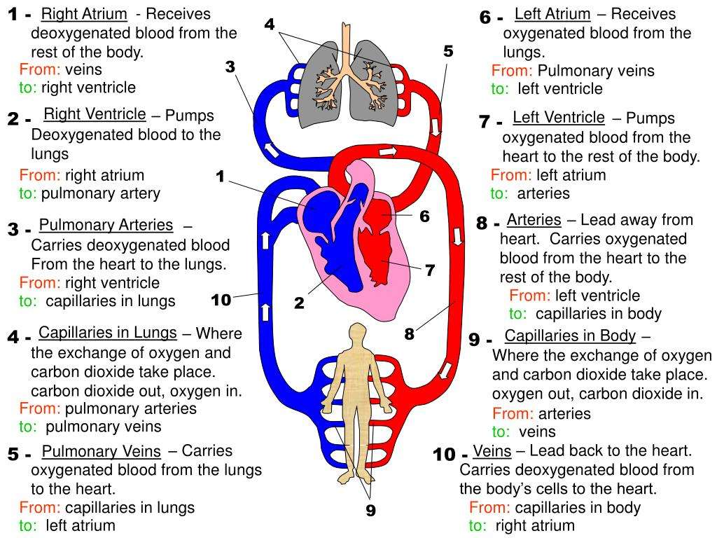
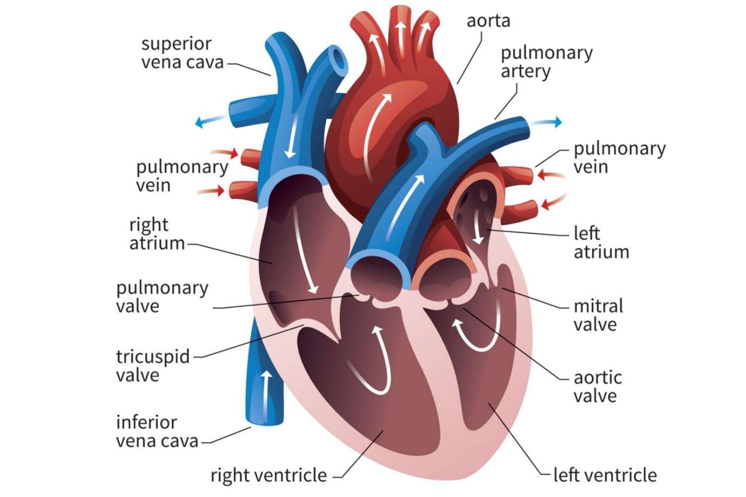
When working properly, deoxygenated blood coming back from organs, other than the lungs, enters the heart through two major veins known as the vena cavae, and the heart returns its venous blood back to itself through the coronary sinus.
From these venous structures, the blood enters the right atrium and passes through the tricuspid valve into the right ventricle. The blood then flows through the pulmonary valve into the pulmonary artery trunk, and next travels through the right and left pulmonary arteries to the lungs, where the blood receives oxygen during air exchange.
On its way back from the lungs, the oxygenated blood travels through the right and left pulmonary veins into the left atrium of the heart. The blood then flows through the mitral valve into the left ventricle, the hearts powerhouse chamber.
The blood travels out the left ventricle through the aortic valve, and into the aorta, extending upward from the heart. From there, the blood moves through a maze of arteries to get to every cell in the body other than the lungs.
Which Of The Following Heart Chambers Pump Oxygenated Blood To The Body
From the left atrium blood flows into the left ventricle. The left ventricle pumps the blood to the aorta which will distribute the oxygenated blood to all parts of the body.
Also to know is, which chamber pumps blood to the body?
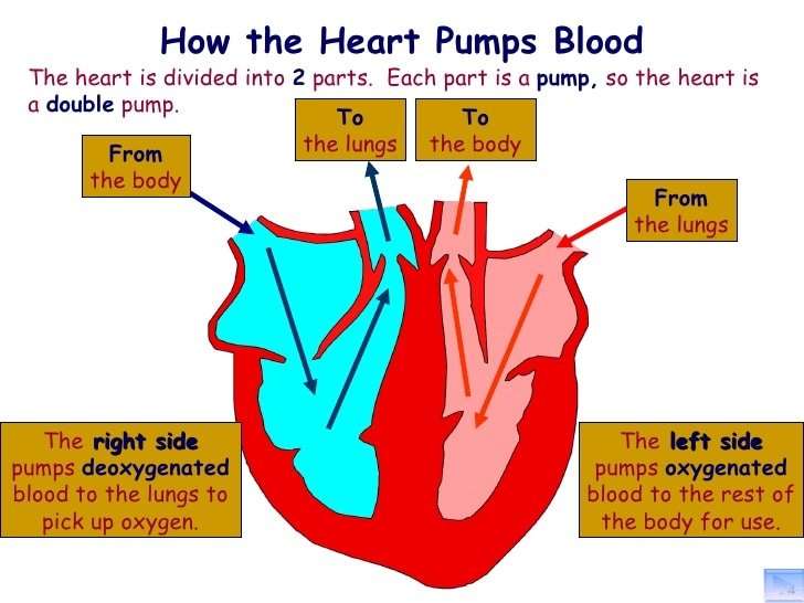
The right ventricle pumps the oxygen-poor blood to the lungs. The left atrium receives oxygen-rich blood from the lungs and pumps it to the left ventricle. The left ventricle pumps the oxygen-rich blood to the body.
Secondly, which heart chamber contains oxygenated blood quizlet? The left atrium, pulmonary veins, and aorta all contain oxygenated blood.
Likewise, which chamber of heart is oxygenated and deoxygenated blood found?
The right and left atria are the top chambers of the heart and receive blood into the heart. The right atrium receives deoxygenated blood from systemic circulation and the left atrium receives oxygenated blood from pulmonary circulation.
Where does the left ventricle pump blood to?
The left ventricle receives oxygenated blood from the left atrium via the mitral valve and pumps it through the aorta via the aortic valve, into the systemic circulation.
You May Like Also
What Is The Correct Order Of The Flow Of Blood
BloodbloodbloodBloodThe heart has four chambers: two atria and two ventricles.
- The right atrium receives oxygen-poor blood from the body and pumps it to the right ventricle.
- The right ventricle pumps the oxygen-poor blood to the lungs.
- The left atrium receives oxygen-rich blood from the lungs and pumps it to the left ventricle.
Recommended Reading: What Causes Low Blood Pressure And High Heart Rate
The Four Chambers Of The Heart
Your heart has a right and left side separated by a wall called the septum. Each side has a small collecting chamber called an atrium, which leads into a large pumping chamber called a ventricle. There are four chambers: the left atrium and right atrium , and the left ventricle and right ventricle .The right side of your heart collects blood on its return from the rest of our body. The blood entering the right side of your heart is low in oxygen. Your heart pumps the blood from the right side of your heart to your lungs so it can receive more oxygen. Once it has received oxygen, the blood returns directly to the left side of your heart, which then pumps it out again to all parts of your body through an artery called the aorta. Blood pressure refers to the amount of force the pumping blood exerts on arterial walls.
The Heart’s Control System

A heartbeat is caused by an electrical impulse traveling through the heart. The heart’s built-in electrical system controls the speed of its pumping. The electrical impulse originates in the sinus node which functions as the heart’s natural pacemaker. The sinus node is most often located in the top of the right atrium. The electrical signals travel through the heart tissue causing the atria and ventricles to contract and relax and the blood to be pumped to the body.
Also Check: Can Acid Reflux Cause Heart Palpitations
The Atria Are The Hearts Entryways For Blood
The left atrium and right atrium are the two upper chambers of the heart. The left atrium receives oxygenated blood from the lungs. The right atrium receives deoxygenated blood returning from other parts of the body. Valves connect the atria to the ventricles, the lower chambers. Each atrium empties into the corresponding ventricle below.
Each Heart Beat Is A Squeeze Of Two Chambers Called Ventricles
The ventricles are the two lower chambers of the heart. Blood empties into each ventricle from the atrium above, and then shoots out to where it needs to go. The right ventricle receives deoxygenated blood from the right atrium, then pumps the blood along to the lungs to get oxygen. The left ventricle receives oxygenated blood from the left atrium, then sends it on to the aorta. The aorta branches into the systemic arterial network that supplies all of the body.
Recommended Reading: Does Tylenol Increase Heart Rate
What Are The Parts Of The Heart
The heart has four chambers two on top and two on bottom:
- The two bottom chambers are the right ventricle and the left ventricle. These pump blood out of the heart. A wall called the interventricular septum is between the two ventricles.
- The two top chambers are the right atrium and the left atrium. They receive the blood entering the heart. A wall called the interatrial septum is between the atria.
The atria are separated from the ventricles by the atrioventricular valves:
- The tricuspid valve separates the right atrium from the right ventricle.
- The mitral valve separates the left atrium from the left ventricle.
Two valves also separate the ventricles from the large blood vessels that carry blood leaving the heart:
- The pulmonic valve is between the right ventricle and the pulmonary artery, which carries blood to the lungs.
- The aortic valve is between the left ventricle and the aorta, which carries blood to the body.
The Flow Of Blood In And Out Of The Heart
Don’t Miss: What Heart Chamber Pushes Blood Through The Aortic Semilunar Valve
Outside View Of The Back Of The Heart
Coronary veins -take oxygen-poor blood that has already been “used” by muscles of the heart and returns it to the right atrium.
Circumflex artery – supplies blood to the left atrium and the side and back of the left ventricle.
Left coronary artery – divides into two branches .
Left anterior descending artery – supplies blood to the front and bottom of the left ventricle and the front of the septum.
Pulmonary veins -bring oxygen-rich blood back to the heart from the lungs.
Right coronary artery – supplies blood to the right atrium, right ventricle, bottom portion of the left ventricle and back of the septum.
Where Does The Aorta Pump Blood To
heartheartarteries
. Accordingly, where does the aorta carry blood to?
The aorta is the large artery that carries oxygen-rich blood from the left ventricle of the heart to other parts of the body.
Furthermore, which area of the aorta supplies blood to the liver? A single celiac trunk emerges and divides into the left gastric artery to supply blood to the stomach and esophagus, the splenic artery to supply blood to the spleen, and the common hepatic artery, which in turn gives rise to the hepatic artery proper to supply blood to the liver, the right gastric artery to
Beside above, which part of the heart pumps blood into the aorta?
Left ventricle: Receives oxygen-rich blood from the left atrium and pumps blood into the aorta. 12. Aortic valve: Allows blood to pass from the left ventricle to the aorta prevents backflow of blood into the left ventricle.
Does the aorta carry oxygenated blood?
The pulmonary veins carry oxygenated blood from the lungs into the left atrium where it is returned to systemic circulation. The aorta is the largest artery in the body. It carries oxygenated blood from the left ventricle of the heart into systemic circulation.
Read Also: Can Acid Reflux Cause Heart Palpitations
How Does Blood Flow Through Your Lungs
Once blood travels through the pulmonic valve, it enters your lungs. This is called the pulmonary circulation. From your pulmonic valve, blood travels to the pulmonary arteries and eventually to tiny capillary vessels in the lungs.
Here, oxygen travels from the tiny air sacs in the lungs, through the walls of the capillaries, into the blood. At the same time, carbon dioxide, a waste product of metabolism, passes from the blood into the air sacs. Carbon dioxide leaves the body when you exhale. Once the blood is oxygenated, it travels back to the left atrium through the pulmonary veins.
How The Heart Works

Your heart is a strong, muscular organ situated slightly to the left of your chest. It pumps blood to all parts of the body through a network of blood vessels by continuously expanding and contracting. On average, your heart will beat 100,000 times and pump about 2,000 gallons of blood each day.
The heart is divided into a right and left side, separated by a septum. Each side has an atrium and a ventricle . The heart has a total of four chambers: right atrium, right ventricle, left atrium and left ventricle.
The right side of the heart collects oxygen-depleted blood and pumps it to the lungs, through the pulmonary arteries, so that the lungs can refresh the blood with a fresh supply of oxygen.
The left side of the heart receives oxygen-rich blood from the lungs, then pumps blood out to the rest of the body’s tissues, through the aorta.
Recommended Reading: How Does Fitbit Calculate Heart Rate
Students Who Viewed This Also Studied
Gargar, Keya_BSMT-1A_Actvitiy#12 _12-01-2020.pdf
Trinity Valley Community College
Gargar, Keya_BSMT-1A_Actvitiy#12 _12-01-2020.pdf
Trinity Valley Community College
Chapter 1 – Chapter 8 Notes
Trinity Valley Community College
Lab Notes – Intro, Microscope, and Cell
Trinity Valley Community College
Trinity Valley Community College BIO 2401
DEVELOS,GARGAR,MEDIODIA, NOVERO, & TOLEDO_Activity 15_BSMT-1A.pdf
Trinity Valley Community College BIO 2401
Gargar, Keya_BSMT-1A_Actvitiy#12 _12-01-2020.pdf
Trinity Valley Community College BIO 2401
Gargar, Keya_BSMT-1A_Actvitiy#12 _12-01-2020.pdf
Trinity Valley Community College BIO 2401
GARGAR, KEYA_BSMT-1A_URINARY PARTICIPATION_12-07-2020.docx
Trinity Valley Community College BIO 2401
Chapter 1 – Chapter 8 Notes
notes
Trinity Valley Community College BIO 2401
Lab Notes – Intro, Microscope, and Cell
notes
Upload your study docs or become a
Course Hero member to access this document
Upload your study docs or become a
Course Hero member to access this document
Electrical Impulses Keep The Beat
The heart’s four chambers pump in an organized manner with the help of electrical impulses that originate in the sinoatrial node . Situated on the wall of the right atrium, this small cluster of specialized cells is the heart’s natural pacemaker, initiating electrical impulses at a normal rate.
The impulse spreads through the walls of the right and left atria, causing them to contract, forcing blood into the ventricles. The impulse then reaches the atrioventricular node, which acts as an electrical bridge for impulses to travel from the atria to the ventricles. From there, a pathway of fibers carries the impulse into the ventricles, which contract and force blood out of the heart.
Don’t Miss: Can Ibs Cause Heart Palpitations
Unlock This Answer Now
Start your 48-hour free trial to unlock this answer and thousands more. Enjoy eNotes ad-free and cancel anytime.
Already a member? Log in here.
The heart is the main organ of the cardiovascular system. The function of the heart is to pump blood. The heart is divided into four chambers: the right and left atrium, and the right and left ventricle. The left side of the heart pumps oxygenated blood and the right side of the heart pumps deoxygenated blood. Blood becomes oxygenated in the lungs and returns to the left atrium via the pulmonary veins. From the left atrium blood is pumped through the bicuspid valve into the left ventricle. From the left ventricle, blood travels to the aorta, which pumps the oxygenated blood to the rest of the body. As blood travels through the body, it dumps off oxygen to tissues and picks up waste products . The now deoxygenated blood returns to the right side of the heart via the vena cava. Deoxygenated blood then travels through the right atrium and to the right ventricle through the tricuspid valve. Deoxygenated blood exits the heart and travels to the lungs through the pulmonary arteries. Once in the lungs, the waste products are removed and oxygen is added to the blood. The blood is now oxygenated and can return to the heart via the pulmonary veins to go through the whole cycle again.
Oxygenated summary
right/left lungs to right/left pulmonary veins to left atrium through bicuspid valve to left ventricle to aorta, to body
Deoxygenated summary
Can You Feel Your Aorta
Youre most likely just feeling your pulse in your abdominal aorta. Your aorta is the main artery that carries blood from your heart to the rest of your body. It runs from your heart, down the center of your chest, and into your abdomen. Its normal to feel blood pumping through this large artery from time to time.
Read Also: Symptoms Of Weak Heart Valves
Valves Maintain Direction Of Blood Flow
As the heart pumps blood, a series of valves open and close tightly. These valves ensure that blood flows in only one direction, preventing backflow.
- The tricuspid valve is situated between the right atrium and right ventricle.
- The pulmonary valve is between the right ventricle and the pulmonary artery.
- The mitral valve is between the left atrium and left ventricle.
- The aortic valve is between the left ventricle and the aorta.
Each heart valve, except for the mitral valve, has three flaps that open and close like gates on a fence. The mitral valve has two valve leaflets.
Left Ventricle Sends Oxygen
The left ventricle relaxes and fills up with blood before squeezing and pumping the oxygen-rich blood through the aortic valve into the aorta the main artery that carries blood to your body. The muscle wall of the left ventricle is very thick because it has to pump blood around the whole body.
You May Like: Constrictive Heart Failure
The Exterior Of The Heart:
Below is a picture of the outside of a normal, healthy, human heart.
The illustration shows the front surface of the heart, including the coronary arteries and major blood vessels.
The heart is the muscle in the lower half of the picture. The heart has four chambers. The right and left atria are shown in purple. The right and left ventricles are shown in red.
Connected to the heart are some of the main blood vessels arteries and veins that make up the blood circulatory system.
The ventricle on the right side of the heart pumps blood from the heart to the lungs.
When you breathe air in, oxygen passes from the lungs through blood vessels where its added to the blood. Carbon dioxide, a waste product, is passed from the blood through blood vessels to the lungs and is removed from the body when you breathe air out.
The atrium on the left side of the heart receives oxygen-rich blood from the lungs.
The pumping action of the left ventricle sends this oxygen-rich blood through the aorta to the rest of the body.
What Are The Parts Of The Circulatory System

Two pathways come from the heart:
- The pulmonary circulation is a short loop from the heart to the lungs and back again.
- The systemic circulation carries blood from the heart to all the other parts of the body and back again.
In pulmonary circulation:
- The pulmonary artery is a big artery that comes from the heart. It splits into two main branches, and brings blood from the heart to the lungs. At the lungs, the blood picks up oxygen and drops off carbon dioxide. The blood then returns to the heart through the pulmonary veins.
In systemic circulation:
Also Check: Does Tylenol Increase Heart Rate
How The Normal Heart Works
The heart is a large muscular organ with the very important job of circulating blood through the blood vessels to the body. Located in the center of the chest, the heart is the hardest working muscle in the human body always working, even while we are sleeping. The heart and blood vessels together make up the body’s cardiovascular system and are vital to supplying the body with the necessary oxygen and nutrients needed to survive. When you breathe, your lungs take in oxygen. The heart pumps blood to the lungs to pick up oxygen, and then it pumps blood through the body to deliver that oxygen.
The animations below show how a normal heart pumps blood. They also explain the changes that happen to a normal heart right after the fetus is born.
