Adding Oxygen To Blood
Oxygen-poor blood from the body enters your heart through two large veins called the superior and inferior vena cava. The blood enters the heart’s right atrium and is pumped to your right ventricle, which in turn pumps the blood to your lungs.
The pulmonary artery then carries the oxygen-poor blood from your heart to the lungs. Your lungs add oxygen to your blood. The oxygen-rich blood returns to your heart through the pulmonary veins. Visit our How the Lungs Work Health Topic to learn more about what happens to the blood in the lungs.
The oxygen-rich blood from the lungs then enters the left atrium and is pumped to the left ventricle. The left ventricle generates the high pressure needed to pump the blood to your whole body through your blood vessels.
When blood leaves the heart to go to the rest of the body, it travels through a large artery called the aorta. A balloon-like bulge, called an aortic aneurysm, can sometimes occur in the aorta.
Improving Health With Current Research
Learn about the following ways the NHLBI continues to translate current research and science into improved health for people who have heart conditions. Research on this topic is part of the NHLBI’s broader commitment to advancing heart and vascular disease scientific discovery.
Learn about some of the pioneering research contributions we have made over the years that have improved clinical care.
Vasculature Of The Head
Blood is carried from your heart to the rest of your body through a complex network of arteries, arterioles, and capillaries. Blood is returned to your heart through venules and veins.
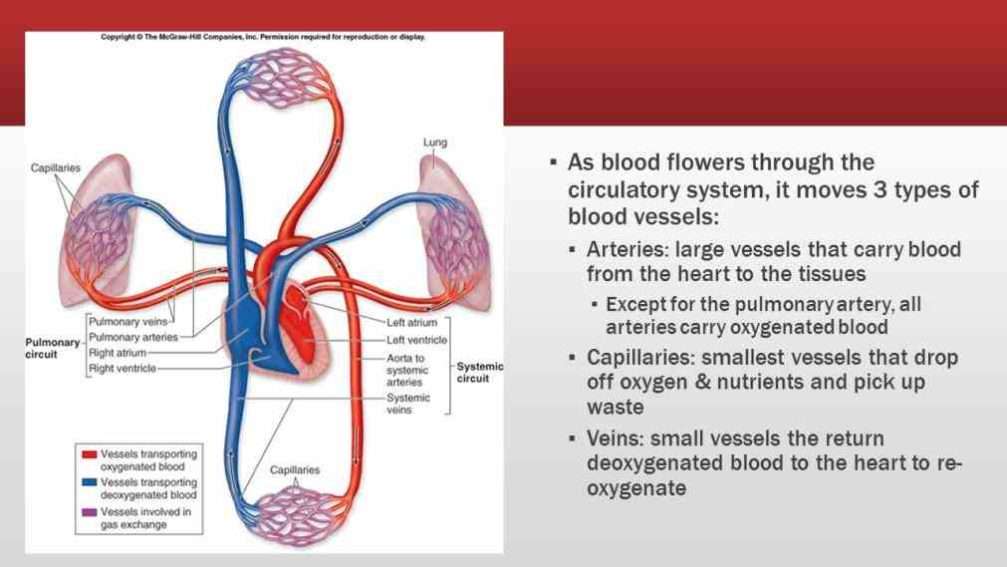
The one-way vascular system carries blood to all parts of your body. This process of blood flow within your body is called circulation. Arteries carry oxygen-rich blood away from your heart, and veins carry oxygen-poor blood back to your heart.
In pulmonary circulation, though, the roles are switched. It is the pulmonary artery that brings oxygen-poor blood into your lungs and the pulmonary vein that brings oxygen-rich blood back to your heart.
In the diagrams, the vessels that carry oxygen-rich blood are colored red, and the vessels that carry oxygen-poor blood are colored blue.
Also Check: Ibs And Heart Palpitations
Anatomy Of Your Heart
Your heart is in the center of your chest, near your lungs. It has four hollow heart chambers surrounded by muscle and other heart tissue. The chambers are separated by heart valves, which make sure that the blood keeps flowing in the right direction. Read more about heart valves in Blood Flow.
Anatomy of the interior of the heart. This image shows the four chambers of the heart and the direction that blood flows through the heart. Oxygen-poor blood, shown in blue-purple, flows into the heart and is pumped out to the lungs. Then oxygen-rich blood, shown in red, is pumped out to the rest of the body, with the help of the heart valves.
Stats About Aortic Aneurysm And Dissection

Impact: According to the CDCs most recent statistics, aortic aneurysms were the main cause of nearly 10,000 deaths in the United States in 2018.
Risk: In 2018, about 58% of people who died from an aortic aneurysm or aortic dissection were male.
Besides advanced age and genetics or family history, people who have the following conditions may be at higher risk for an aortic aneurysm or dissection:
- High blood pressure. The increased force of blood can weaken the artery walls.
- Genetic conditions, such as Marfans Syndrome, decrease the bodys ability to make healthy connective tissue.
- High cholesterol or atherosclerosis. A build-up of plaque may cause increased inflammation in and around the aorta and other blood vessels.
- Inflamed arteries. Certain diseases and conditions, such as vasculitis, can cause the bodys blood vessels to become inflamed.
- Trauma, such as car accidents.
- Smoking. People with a history of smoking make up 75% of abdominal aortic aneurysms.
Screening: The U.S. Preventive Services Task Force recommends that men ages 65-75 who have ever smoked get an ultrasound screening for abdominal aortic aneurysms, even if they have no symptoms.
People living with aortic disease or other heart-related conditions can improve their odds of living a longer and healthier life. Its important to:
- Report any symptoms immediately.
Recommended Reading: Does Benadryl Lower Heart Rate
How Blood Flows Through The Body
As the heart pumps, blood is pushed through the body through the entire circulatory system. Oxygenated blood is pumped away from the heart to the rest of the body, while deoxygenated blood is pumped to the lungs where it is reoxygenated before returning to the heart.
Circulatory system: This illustration of the circulatory system shows where blood flows in the body. Red indicates oxygenated blood, while blue indicates deoxygenated blood.
What Is The Difference Between An Artery And A Vein
- carry blood from the tissues of the body back to the heart
- are usually positioned closer beneath the surface of the skin
- are less muscular than arteries, but contain valves to help keep blood flowing in the right direction, usually toward the heart
- would collapse if blood flow stops.
- carry blood away from the heart to the tissues of the body
- are usually positioned deeper within the body
- are more muscular than veins, which helps in transporting blood that is full of oxygen efficiently to the tissues
- would generally remain open if blood flow stopped, due to their thick muscular layer.
You may have been told that you had a heart attack or a stroke because of a blocked artery. What exactly is an artery?
Arteries, like veins, are tube-shaped vessels that carry blood in the body. The chief difference between arteries and veins is the job that they do. Arteries carry oxygenated blood away from the heart to the body, and veins carry oxygen-poor blood back from the body to the heart.
Your body also contains other, smaller blood vessels. Here is how blood travels in vessels through the body:
Recommended Reading: Can Ibs Cause Heart Palpitations
What Are The Different Types Of Arteries
There are three types of arteries. Each type is composed of three coats: outer, middle, and inner.
- Elastic arteries are also called conducting arteries or conduit arteries. They have a thick middle layer so they can stretch in response to each pulse of the heart.
- Muscular arteries are medium-sized. They draw blood from elastic arteries and branch into resistance vessels. These vessels include small arteries and arterioles.
- Arterioles are the smallest division of arteries that transport blood away from the heart. They direct blood into the capillary networks.
There are four types of veins:
- Deep veins are located within muscle tissue. They have a corresponding artery nearby.
- Superficial veins are closer to the skins surface. They dont have corresponding arteries.
- Pulmonary veins transport blood thats been filled with oxygen by the lungs to the heart. Each lung has two sets of pulmonary veins, a right and left one.
- Systemic veins are located throughout the body from the legs up to the neck, including the arms and trunk. They transport deoxygenated blood back to the heart.
Use this interactive 3-D diagram to explore an artery.
Use this interactive 3-D diagram to explore a vein.
How Do The Heart And Blood Vessels Work
The heart works by following a sequence of electrical signals that cause the muscles in the chambers of the heart to contract in a certain order. If these electrical signals change, the heart may not pump as well as it should.
The sequence of each heartbeat is as follows:
- The sinoatrial node in the right atrium is like a tiny in-built ‘timer’. It fires off an electrical impulse at regular intervals. This controls your heart rate. Each impulse spreads across both atria, which causes them to contract. This pumps blood through one-way valves into the ventricles.
- The electrical impulse gets to the atrioventricular node at the lower right atrium. This acts like a ‘junction box’ and the impulse is delayed slightly. Most of the tissue between the atria and ventricles does not conduct the impulse. However, a thin band of conducting fibres called the atrioventricular bundle acts like ‘wires’ and carries the impulse from the AV node to the ventricles.
- The AV bundle splits into two – a right and a left branch. These then split into many tiny fibres which carry the electrical impulse throughout the ventricles. The ventricles contract and pump blood through one-way valves into large arteries:
- The arteries going from the right ventricle take blood to the lungs.
- The arteries going from the left ventricle take blood to the rest of the body.
Don’t Miss: Flonase Heart Racing
What Are The Parts Of The Heart
The heart has four chambers two on top and two on bottom:
- The two bottom chambers are the right ventricle and the left ventricle. These pump blood out of the heart. A wall called the interventricular septum is between the two ventricles.
- The two top chambers are the right atrium and the left atrium. They receive the blood entering the heart. A wall called the interatrial septum is between the atria.
The atria are separated from the ventricles by the atrioventricular valves:
- The tricuspid valve separates the right atrium from the right ventricle.
- The mitral valve separates the left atrium from the left ventricle.
Two valves also separate the ventricles from the large blood vessels that carry blood leaving the heart:
- The pulmonic valve is between the right ventricle and the pulmonary artery, which carries blood to the lungs.
- The aortic valve is between the left ventricle and the aorta, which carries blood to the body.
Blood Vessels: Circulating The Blood
|
Blood travels from the heart in arteries, which branch into smaller and smaller vessels, eventually becoming arterioles. Arterioles connect with even smaller blood vessels called capillaries. Through the thin walls of the capillaries, oxygen and nutrients pass from blood into tissues, and waste products pass from tissues into blood. From the capillaries, blood passes into venules, then into veins to return to the heart. Arteries and arterioles have relatively thick muscular walls because blood pressure in them is high and because they must adjust their diameter to maintain blood pressure and to control blood flow. Veins and venules have much thinner, less muscular walls than arteries and arterioles, largely because the pressure in veins and venules is much lower. Veins may dilate to accommodate increased blood volume. |
If a blood vessel breaks, tears, or is cut, blood leaks out, causing bleeding. Blood may flow out of the body, as external bleeding, or it may flow into the spaces around organs or directly into organs, as internal bleeding.
Don’t Miss: Ibs Heart Palpitations
Anatomy Of Veins And Arteries
The walls of veins and arteries are both made up of three layers:
- Outer. Tunica adventitia is the outer layer of a blood vessel, including arteries and veins. Its mostly composed of collagen and elastic fibers. These fibers enable the veins and arteries to stretch a limited amount. They stretch enough to be flexible while maintaining stability under the pressure of blood flow.
- Middle. The middle layer of the walls of arteries and veins is called the tunica media. Its made of smooth muscle and elastic fibers. This layer is thicker in arteries and thinner in veins.
- Inner. The inner layer of the blood vessel wall is called tunica intima. This layer is made of elastic fiber and collagen. Its consistency varies based on the type of blood vessel.
Unlike arteries, veins contain valves. Veins need valves to keep the blood flowing toward the heart. Theses valves are particularly important in the legs and arms. They fight gravity to prevent the backflow of blood.
Arteries dont need valves because the pressure from the heart keeps the blood flowing through them in one direction.
Heart Anatomy And Circulation

The heart is located in the thoracic cavity in a central compartment of the cavity known as the mediastinum. It is situated between the left and right lungs in the chest cavity. The heart is divided into upper and lower chambers called atria and ventricles . These chambers function to collect blood returning to the heart from circulation and to pump blood out of the heart. The heart is a major structure of the cardiovascular system as it serves to drive blood to all the cells of the body. Blood is circulated along a pulmonary circuit and a systemic circuit. The pulmonary circuit involves the transport of blood between the heart and lungs, while the systemic circuit involves blood circulation between the heart and the rest of the body.
Don’t Miss: Can Flonase Cause Heart Palpitations
Blood Vessels Leading Into And Out Of The Heart
There are four main blood vessels that take blood into and out of the heart.
Arteries carry oxygenated blood away from the heart .
The main artery is the aorta.
The main vein is the vena cava.
Pulmonary Trunk And Pulmonary Arteries
The main pulmonary artery or pulmonary trunk is a part of the pulmonary circuit. It is a large artery and one of the three major blood vessels that extend from the heart. The other major vessels include the aorta and vena cavae. The pulmonary trunk is connected to the right ventricle of the heart and receives oxygen-poor blood. The pulmonary valve, located near the opening of the pulmonary trunk, prevents blood from flowing back into the right ventricle. Blood is conveyed from the pulmonary trunk to the left and right pulmonary arteries.
Don’t Miss: How Do You Say Heart Attack In Spanish
Types Of Arteries And Veins
There are two types of arteries in the body: Pulmonary and systemic. The pulmonary artery carries deoxygenated blood from the heart, to the lungs, for purification while the systemic arteries form a network of arteries that carry oxygenated blood from the heart to other parts of the body. Arterioles and capillaries are further extensions of the artery that help transport blood to tinier parts in the body.
Veins can be classified as pulmonary veins and systemic veins. The pulmonary veins are a set of veins that deliver oxygenated blood from the lungs to the heart and the Systemic veins drain the tissues of the body and deliver deoxygenated blood to the heart. Pulmonary and Systemic veins can either be superficial or embedded deep inside the body.
What Causes Vascular Disease
Causes of vascular disease include:
-
Atherosclerosis. Atherosclerosis is the most common cause of vascular disease. It is unknown exactly how atherosclerosis starts or what causes it. Atherosclerosis is a slow, progressive, vascular disease that may start as early as childhood. However, the disease has the potential to progress rapidly. It is generally characterized by the buildup of fatty deposits along the innermost layer of the arteries. If the disease process progresses, plaque may form. This thickening narrows the arteries and can decrease blood flow or completely block the flow of blood to organs and other body tissues and structures.
-
Blood clots. A blood vessel may be blocked by an embolus or a thrombus .
-
Inflammation. In general, inflammation of blood vessels is referred to as vasculitis, which includes a range of disorders. Inflammation may lead to narrowing and blockage of blood vessels.
-
Trauma or injury. Trauma or injury involving the blood vessels may lead to inflammation or infection, which can damage the blood vessels and lead to narrowing and blockage.
-
Genetic. Certain conditions of the vascular system are inherited.
Recommended Reading: Does Benadryl Lower Heart Rate
Exchange Of Gases Nutrients And Waste Between Blood And Tissue Occurs In The Capillaries
Capillaries are tiny vessels that branch out from arterioles to form networks around body cells. In the lungs, capillaries absorb oxygen from inhaled air into the bloodstream and release carbon dioxide for exhalation. Elsewhere in the body, oxygen and other nutrients diffuse from blood in the capillaries to the tissues they supply. The capillaries absorb carbon dioxide and other waste products from the tissues and then flow the deoxygenated blood into the veins.
The Blood Supply To The Heart
Like any other muscle, the heart muscle needs a good blood supply. The coronary arteries take blood to the heart muscle. These are the first arteries to branch off the large artery which takes blood to the body from the left ventricle.
- The right coronary artery mainly supplies the muscle of the right ventricle.
- The left coronary artery quickly splits into two and supplies the rest of the heart muscle.
- The main coronary arteries divide into many smaller branches to supply all the heart muscle.
Read Also: Does Benadryl Lower Heart Rate
What Are The Heart And Blood Vessels
Blood vessels form the living system of tubes that carry blood both to and from the heart. All cells in the body need oxygen and the vital nutrients found in blood. Without oxygen and these nutrients, the cells will die. The heart helps to provide oxygen and nutrients to the body’s tissues and organs by ensuring a rich supply of blood.
Not only do blood vessels carry oxygen and nutrients, they also transport carbon dioxide and waste products away from our cells. Carbon dioxide is passed out of the body by the lungs most of the other waste products are disposed of by the kidneys. Blood also transports heat around your body.
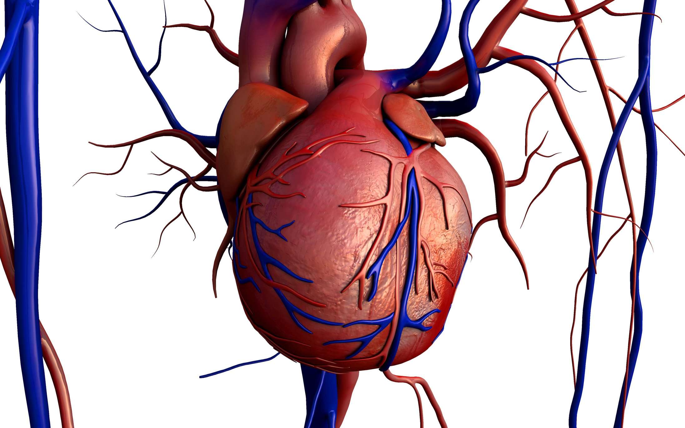
Anatomy of the heart and blood vessels
