How The Heart Works
The heart is an organ, about the size of a fist. It is made of muscle and pumps blood through the body. Blood is carried through the body in blood vessels, or tubes, called arteries and veins. The process of moving blood through the body is called circulation. Together, the heart and vessels make up the cardiovascular system.
Blood Vessels Of The Heart
The blood vessels of the heart include:
- venae cavae deoxygenated blood is delivered to the right atrium by these two veins. One carries blood from the head and upper torso, while the other carries blood from the lower body
- pulmonary arteries deoxygenated blood is pumped by the right ventricle into the pulmonary arteries that link to the lungs
- pulmonary veins the pulmonary veins return oxygenated blood from the lungs to the left atrium of the heart
- aorta this is the largest artery of the body, and it runs the length of the trunk. Oxygenated blood is pumped into the aorta from the left ventricle. The aorta subdivides into various branches that deliver blood to the upper body, trunk and lower body
- coronary arteries like any other organ or tissue, the heart needs oxygen. The coronary arteries that supply the heart are connected directly to the aorta, which carries a rich supply of oxygenated blood
- coronary veins deoxygenated blood from heart muscle is ‘dumped’ by coronary veins directly into the right atrium.
Chambers Of The Heart
The internalcavity of the heart is divided into four chambers:
- Right atrium
- Left atrium
- Left ventricle
The two atria are thin-walled chambers that receive blood from the veins. The two ventricles are thick-walled chambers that forcefully pump blood out of the heart. Differences in thickness of the heart chamber walls are due to variations in the amount of myocardium present, which reflects the amount of force each chamber is required to generate.
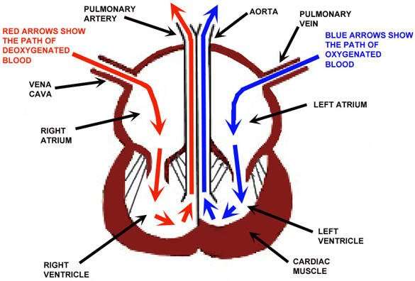
The right atrium receives deoxygenated blood from systemic veins the left atrium receives oxygenated blood from the pulmonary veins.
Don’t Miss: Flonase And Heart Palpitations
The Atria Are The Hearts Entryways For Blood
The left atrium and right atrium are the two upper chambers of the heart. The left atrium receives oxygenated blood from the lungs. The right atrium receives deoxygenated blood returning from other parts of the body. Valves connect the atria to the ventricles, the lower chambers. Each atrium empties into the corresponding ventricle below.
The Heart Wall Is Composed Of Three Layers

The muscular wall of the heart has three layers. The outermost layer is the epicardium . The epicardium covers the heart, wraps around the roots of the great blood vessels, and adheres the heart wall to a protective sac. The middle layer is the myocardium. This strong muscle tissue powers the hearts pumping action. The innermost layer, the endocardium, lines the interior structures of the heart.
Recommended Reading: Fitbit Charge 2 Accuracy Heart Rate
The Interior Of The Heart
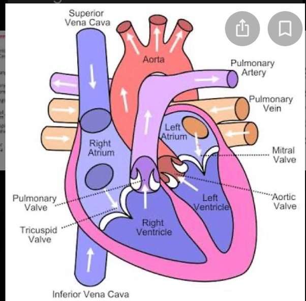
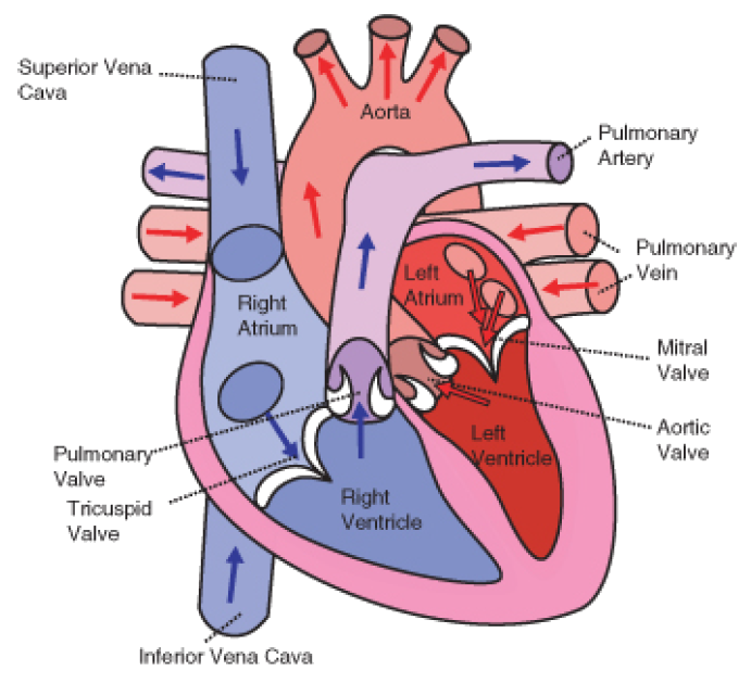
Below is a picture of the inside of a normal, healthy, human heart.
The illustration shows a cross-section of a healthy heart and its inside structures. The blue arrow shows the direction in which low-oxygen blood flows from the body to the lungs. The red arrow shows the direction in which oxygen-rich blood flows from the lungs to the rest of the body.
The Valves Are Like Doors To The Chambers Of The Heart
Four valves regulate and support the flow of blood through and out of the heart. The blood can only flow one waylike a car that must always be kept in drive. Each valve is formed by a group of folds, or cusps, that open and close as the heart contracts and dilates. There are two atrioventricular valves, located between the atrium and the ventricle on either side of the heart: The tricuspid valve on the right has three cusps, the mitral valve on the left has two. The other two valves regulate blood flow out of the heart. The aortic valve manages blood flow from the left ventricle into the aorta. The pulmonary valve manages blood flow out of the right ventricle through the pulmonary trunk into the pulmonary arteries.
You May Like: Can Flonase Cause Heart Palpitations
The Right Side Of The Heart
The superior and inferior vena cavae are in blue to the left of the muscle as you look at the picture. These veins are the largest veins in the body. They carry used blood to the right atrium of the heart.
Used blood has had its oxygen removed and used by the bodys organs and tissues. The superior vena cava carries used blood from the upper parts of the body, including the head, chest, arms, and neck. The inferior vena cava carries used blood from the lower parts of the body.
The used blood from the vena cavae flows into the hearts right atrium and then on to the right ventricle. From the right ventricle, the used blood is pumped through the pulmonary arteries to the lungs. Here, through many small, thin blood vessels called capillaries, the blood picks up oxygen needed by all the areas of the body.
The oxygen-rich blood passes from the lungs back to the heart through the pulmonary veins .
Which Side Of The Heart Contain Oxygenated Blood
Oxygenated blood returns to the LEFT side of the heart, into the left atrium, down into the left ventricle and out into the aorta to perfuse the tissues of the body.
Step-by-step explanation:
Because blood is pumped to the lungs, where we can find fresh blood. The blood in the RIGHT side of the heart is deoxygenated. The left side of the heart is the one which receives oxygenated blood. It comes directly from the pulmonary vein. It is further pumped into the aorta. The right side of the heart receives deoxygenated blood from the phenomenon called vena cava. Then, its pumped into the pulmonary vein.
Also Check: What Heart Chamber Pushes Blood Through The Aortic Semilunar Valve
What Are The Parts Of The Heart
The heart has four chambers two on top and two on bottom:
- The two bottom chambers are the right ventricle and the left ventricle. These pump blood out of the heart. A wall called the interventricular septum is between the two ventricles.
- The two top chambers are the right atrium and the left atrium. They receive the blood entering the heart. A wall called the interatrial septum is between the atria.
The atria are separated from the ventricles by the atrioventricular valves:
- The tricuspid valve separates the right atrium from the right ventricle.
- The mitral valve separates the left atrium from the left ventricle.
Two valves also separate the ventricles from the large blood vessels that carry blood leaving the heart:
- The pulmonic valve is between the right ventricle and the pulmonary artery, which carries blood to the lungs.
- The aortic valve is between the left ventricle and the aorta, which carries blood to the body.
What Are The Congenital Heart Defects That Involve Mismatched Blood Vessels Near The Heart
The location and formation of the blood vessels that connect with the heart are important, because the location determines the type of blood that a vessel receives. If the body receives deoxygenated blood or the lungs receive oxygenated blood, the heart will be strained or unable to meet oxygen demands in the body.
Transposition of the great arteries is a congenital heart disease in which the aorta and pulmonary artery have been mismatched in their connection to the heart. Usually, the aorta receives oxygenated blood from the left ventricle and delivers it to the body. But in a patient with transposition of the great arteries, the aorta receives blood that is poor in oxygen from the right ventricle and carries this blood to the body, and the pulmonary artery receives oxygen-rich blood from the left ventricle to be cycled again through the lungs. Thus, without some sort of communication between the two sides, the same blood is continually pumped through the body and lungs.
Truncus arteriosus is a congenital defect in which the aorta and the pulmonary artery fail to form separately, but are joined permanently as they rise from the heart and separate as they branch into smaller arteries that deliver blood to the lungs and body. This makes it impossible for the heart to segregate the oxygenated and deoxygenated blood that comes from the two sides of the heart. Truncus arteriosus makes a patients heart inefficient because it delivers the same blood to the lungs and body.
Recommended Reading: 10 Second Trick To Prevent Heart Attack
What Side Of The Heart Receives Oxygenated Blood
leftright
The right ventricle pumps the blood to the lungs where it becomes oxygenated. The oxygenated blood is brought back to the heart by the pulmonary veins which enter the left atrium. The left ventricle pumps the blood to the aorta which will distribute the oxygenated blood to all parts of the body.
Subsequently, question is, why is the right side of the heart deoxygenated? The heart is a unidirectional pump. Valves are present to prevent the backflow of blood. The right side pumps deoxygenated blood to the lungs. The left side pumps oxygenated blood to the organs of the body.
Besides, is the left ventricle oxygenated or deoxygenated?
The right ventricle receives deoxygenated blood from the right atrium, then pumps the blood along to the lungs to get oxygen. The left ventricle receives oxygenated blood from the left atrium, then sends it on to the aorta. The aorta branches into the systemic arterial network that supplies all of the body.
Which side of heart has oxygen rich blood?
The right side of the heart pumps oxygen-poor blood from the body to the lungs, where it receives oxygen. The left side of the heart pumps oxygen-rich blood from the lungs to the body.
How Does Blood Flow Through Your Lungs

Once blood travels through the pulmonic valve, it enters your lungs. This is called the pulmonary circulation. From your pulmonic valve, blood travels to the pulmonary artery to tiny capillary vessels in the lungs. Here, oxygen travels from the tiny air sacs in the lungs, through the walls of the capillaries, into the blood. At the same time, carbon dioxide, a waste product of metabolism, passes from the blood into the air sacs. Carbon dioxide leaves the body when you exhale. Once the blood is purified and oxygenated, it travels back to the left atrium through the pulmonary veins.
You May Like: Can Benadryl Cause Arrhythmias
How Do Doctors Treat Congenital Heart Defects That Involve Mismatched Blood Vessels Near The Heart
Transposition of the great arteries requires immediate intervention to get adequate oxygen to the body for vital functions. If the oxygenated and deoxygenated blood is kept completely separate, a person cannot survive long. If, however, a septal defect or patent ductus exists in the heart or arteries, the two blood supplies will be able to communicate, and a patient may be able to retain vital functions. Balloon atrial septostomy, or the Rashkind procedure, is a catheter-based procedure that doctors can use to widen a hole in the atrial or ventricular walls in order to allow better communication between the two bloods. A surgeon may perform one of two procedures to correct transposition of the great arteries, venous switch or arterial switch. Both of these procedures try to create a situation conducive to normal blood flow by switching the connections.
Patients with the tetralogy of Fallot may need a procedure to allow blood to mix between the aorta and the pulmonary artery in order to survive infancy. In early childhood, most patients with the tetralogy of Fallot will need open heart surgery to close the ventricular septal defect and widen the stenosis in the pulmonary valve.
Most people born with truncus arteriosus will need a surgical procedure in which a surgeon separates the two arteries and makes connections between the pulmonary artery and the right ventricle.
Each Heart Beat Is A Squeeze Of Two Chambers Called Ventricles
The ventricles are the two lower chambers of the heart. Blood empties into each ventricle from the atrium above, and then shoots out to where it needs to go. The right ventricle receives deoxygenated blood from the right atrium, then pumps the blood along to the lungs to get oxygen. The left ventricle receives oxygenated blood from the left atrium, then sends it on to the aorta. The aorta branches into the systemic arterial network that supplies all of the body.
Recommended Reading: Ibs And Heart Palpitations
How Do Doctors Treat Septal Defects And Patent Ductus Arteriosus
Surgeons can sew a patch in the septal wall of patients with atrial and ventricular septal defects to ensure normal blood flow in the heart. A less invasive alternative to heart surgery is a catheter based procedure in which a doctor places a small, expandable disk in the hole through a small tube inserted through a blood vessel somewhere else in the body and threaded to the heart. A small hole may not need treatment because it may heal itself or cause only an insignificant decrease in heart efficiency. Sometimes, drug therapy can help to close the connection between the arteries in patent ductus arteriosus, but surgery may be necessary to fix the defect.
What Are Congenital Heart Defects
Congenital heart defects are problems with the hearts structure that developed in the womb or early development and are present at birth or shortly thereafter. Each year, about 1 percent or 30,000 babies are born with the defect, making it the most common birth defect in the U. S. Congenital heart defects vary widely in structure and severity. Some defects can be fatal, but thanks largely to new treatments, most affected individuals survive their childhood and live relatively normal lives. Over 1,000,000 adults are currently living with a congenital heart disease.
Don’t Miss: Will Benadryl Help Heart Palpitations
Heart Anatomy: By The Numbers
1. Superior vena cava: Receives blood from the upper body delivers blood into the right atrium.
2. Inferior vena cava: Receives blood from the lower extremities, pelvis and abdomen, and delivers blood into the right atrium.
3. Right atrium: Receives blood returning to the heart from the superior and inferior vena cava transmits blood to the right ventricle, which pumps blood to the lungs for oxygenation.
4. Tricuspid valve: Allows blood to pass from the right atrium to the right ventricle prevents blood from flowing back into the right atrium as the heart pumps .
5. Right ventricle: Receives blood from the right atrium pumps blood into the pulmonary artery.
6. Pulmonary valve: Allows blood to pass into the pulmonary arteries prevents blood from flowing back into the right ventricle.
7. Pulmonary arteries: Carry oxygen-depleted blood from the heart to the lungs.
8. Pulmonary veins: Deliver oxygen-rich blood from the lungs to the left atrium of the heart.
9. Left atrium: Receives blood returning to the heart from the pulmonary veins.
10. Mitral valve: Allows blood to flow into the left ventricle prevents blood from flowing back into the left atrium.
11. Left ventricle: Receives oxygen-rich blood from the left atrium and pumps blood into the aorta.
12. Aortic valve: Allows blood to pass from the left ventricle to the aorta prevents backflow of blood into the left ventricle.
13. Aorta: Distributes blood throughout the body from the heart.
This Problem Has Been Solved
1. What is the relationship between blood pressure, heart rate,and respiratory rate when there is blood loss? Explain.
2. Explain systemic cardiac output. What factors areinvolved in cardiac output?
3. Which side of the heart contains oxygenated blood?Explain. Which side of the heart contains deoxygenated blood?Explain.
4. Trace the path of a blood cell as it travels through thebody. Start and end with the inferior vena cava. = Then, trace thepath of a blood cell traveling through the body starting and endingwith the superior vena cava.
Please answer with detail
You May Like: Can Flonase Cause Heart Palpitations
What Does The Circulatory System Do
The circulatory system is made up of blood vessels that carry blood away from and towards the heart. Arteries carry blood away from the heart and veins carry blood back to the heart.
The circulatory system carries oxygen, nutrients, and to cells, and removes waste products, like carbon dioxide. These roadways travel in one direction only, to keep things going where they should.
The Cardiac Cycle Includes All Blood Flow Events The Heart Accomplishes In One Complete Heartbeat

The muscular wall of the heart powers contraction and dilation. Each contraction and relaxation is a heartbeat. Ventricular contractions, called systole, force blood out of the heart through the pulmonary and aortic valves. Diastole occurs when blood flows from the atria to fill the ventricles.
You May Like: What Is A Typical Resting Heart Rate For A Healthy Individual
How Do Doctors Identify Congenital Heart Defects
Some heart defects can be seen on prenatal tests and the doctor can order a fetal echocardiogram if he suspects a defect in the child. Many defects, however, are identified shortly after birth by their signs and symptoms. Some signs and symptoms of defects include a heart murmur a bluish tint to skin, lips, and fingernails shortness of breath fast breathing fatigue and poor weight gain. If a doctor suspects a heart defect in a newborn, he may order an echocardiogram, an EKG, a chest x-ray, or a pulse oximetry. A pulse oximetry is a noninvasive test doctors use to see if oxygenated and deoxygenated blood is mixing in the heart and to observe lung function. The test involves placing a sensor similar to a bandage on the childs finger or toe. A cardiac catheterization may be necessary to definitively see the arteries around the heart and observe heart function.
Please be aware that this information is provided to supplement the care provided by your physician. It is neither intended nor implied to be a substitute for professional medical advice. CALL YOUR HEALTHCARE PROVIDER IMMEDIATELY IF YOU THINK YOU MAY HAVE A MEDICAL EMERGENCY. Always seek the advice of your physician or other qualified health provider prior to starting any new treatment or with any questions you may have regarding a medical condition.
