What Are The Different Types Of Arteries
Arteries are the blood vessels that carry blood away from the heart. The different types of arteries include:
- Elastic arteries: are also called conducting arteries that have a thick middle layer that stretches in response to each pulse.
- Muscular arteries: are medium-sized arteries that draw blood from elastic arteries.
- Arterioles direct the blood into the capillaries and are arterial divisions that transport blood away from the heart.
Coronary Artery Disease : Coronary Occlusion
Coronary arteries are end arteries.
If it gets blocked, then there is no other way the blood could reach other parts of the heart, beyond the block.
If there is a plaque blocking a coronary artery, then it would result in heart attack, which means, the areas supplied beyond the block undergo ischemic changes , which lead to death of portion of the heart.
The extent of damage to the heart depends on the degree of the block, which could be found out by Angiography.
The degree of block is expressed in percentage of blocked lumen , if its 100% block then the blood cannot pass through it, while partial blocks allow a limited portion but still compromising the vital blood supply.
Irasto Medical TeamReviewed on 23 May, 2021.
Venous Drainage Of The Brain
Circulation to the brain is both critical and complex. Many smaller veins of the brain stem and the superficial veins of the cerebrum lead to larger vessels referred to as intracranial sinuses. These include the superior and inferior sagittal sinuses, straight sinus, cavernous sinuses, left and right sinuses, the petrosal sinuses, and the occipital sinuses. Ultimately, sinuses will lead back to either the inferior jugular vein or vertebral vein.
Figure 15. This left lateral view shows the veins of the head and neck, including the intercranial sinuses.
| Table 11. Major Veins of the Brain | |
|---|---|
| Vessel | |
| Enlarged vein that receives blood from the cavernous sinus and leads into the internal jugular veins | |
| Occipital sinus | Enlarged vein that drains the occipital region near the falx cerebelli and leads to the left and right transverse sinuses, and also the vertebral veins |
| Transverse sinuses | Pair of enlarged veins near the lambdoid suture that drains the occipital, sagittal, and straight sinuses, and leads to the sigmoid sinuses |
| Sigmoid sinuses | Enlarged vein that receives blood from the transverse sinuses and leads through the jugular foramen to the internal jugular vein |
Don’t Miss: Why Do Av Nodal Cells Not Determine The Heart Rate
Classification & Structure Of Blood Vessels
Blood vessels are the channels or conduits through which blood is distributed to body tissues. The vessels make up two closed systems of tubes that begin and end at the heart. One system, the pulmonary vessels, transports blood from the right ventricle to the lungs and back to the left atrium. The other system, the systemic vessels, carries blood from the left ventricle to the tissues in all parts of the body and then returns the blood to the right atrium. Based on their structure and function, blood vessels are classified as either arteries, capillaries, or veins.
Also Check: What Do You Do If Someone Is Having A Heart Attack
What Do The Heart And Blood Vessels Do

The heart’s main function is to pump blood around the body. Blood carries nutrients and waste products and is vital to life. One of the essential nutrients found in blood is oxygen.
The right side of the heart receives blood lacking oxygen from the body. After passing through the right atrium and right ventricle this blood is pumped to the lungs. Here blood picks up oxygen and loses another gas called carbon dioxide. Once through the lungs, the blood flows back to the left atrium. It then passes into the left ventricle and is pumped into the main artery supplying the body. Oxygenated blood is then carried though blood vessels to all the body’s tissues. Here oxygen and other nutrients pass into the cells where they are used to perform the body’s essential functions.
A blood vessel’s main function is to transport blood around the body. Blood vessels also play a role in controlling your blood pressure.
Blood vessels are found throughout the body. There are five main types of blood vessels: arteries, arterioles, capillaries, venules and veins.
Arteries carry blood away from the heart to other organs. They can vary in size. The largest arteries have special elastic fibres in their walls. This helps to complement the work of the heart, by squeezing blood along when heart muscle relaxes. Arteries also respond to signals from our nervous system, either tightening or relaxing .
Read Also: Can Ibs Cause Heart Palpitations
Anatomy Of The Heart And Blood Vessels
Reviewed byDr Jacqueline Payne
The heart is a muscular pump that pushes blood through blood vessels around the body. The heart beats continuously, pumping the equivalent of more than 14,000 litres of blood every day through five main types of blood vessels: arteries, arterioles, capillaries, venules and veins.
Read Also: Is 116 Heart Rate High
What Is Collateral Circulation
Collateral circulation is a network of tiny blood vessels, and, under normal conditions, not open. When the coronary arteries narrow to the point that blood flow to the heart muscle is limited , collateral vessels may enlarge and become active. This allows blood to flow around the blocked artery to another artery nearby or to the same artery past the blockage, protecting the heart tissue from injury.
Collateral vessels surround blocked blood vessel
Also Check: Can Flonase Cause Heart Palpitations
Describe Briefly The Cardiac Dominance
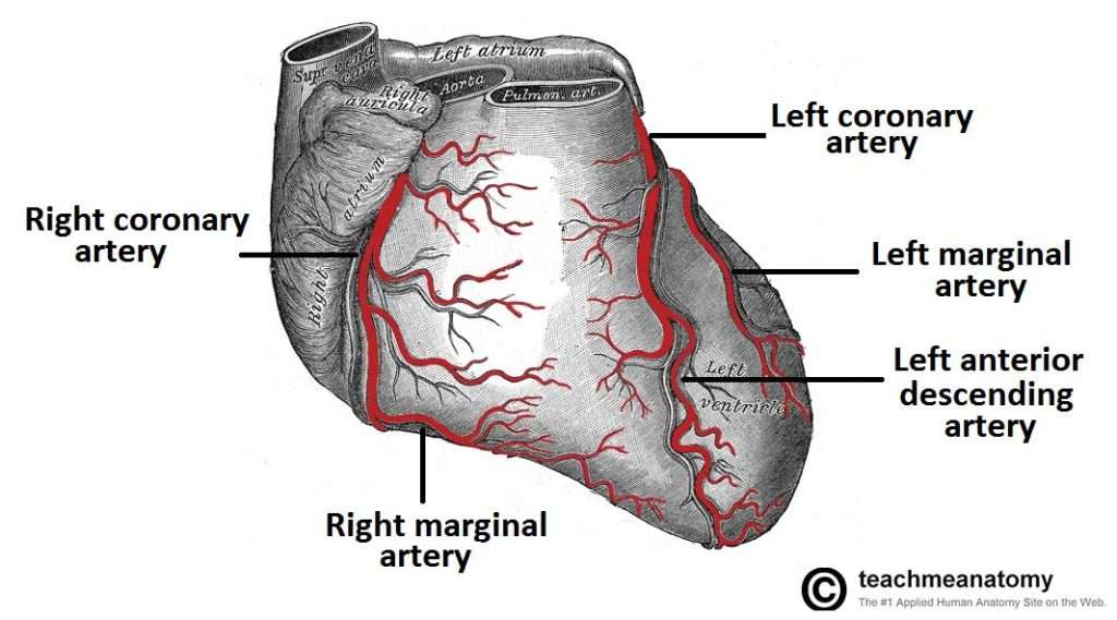
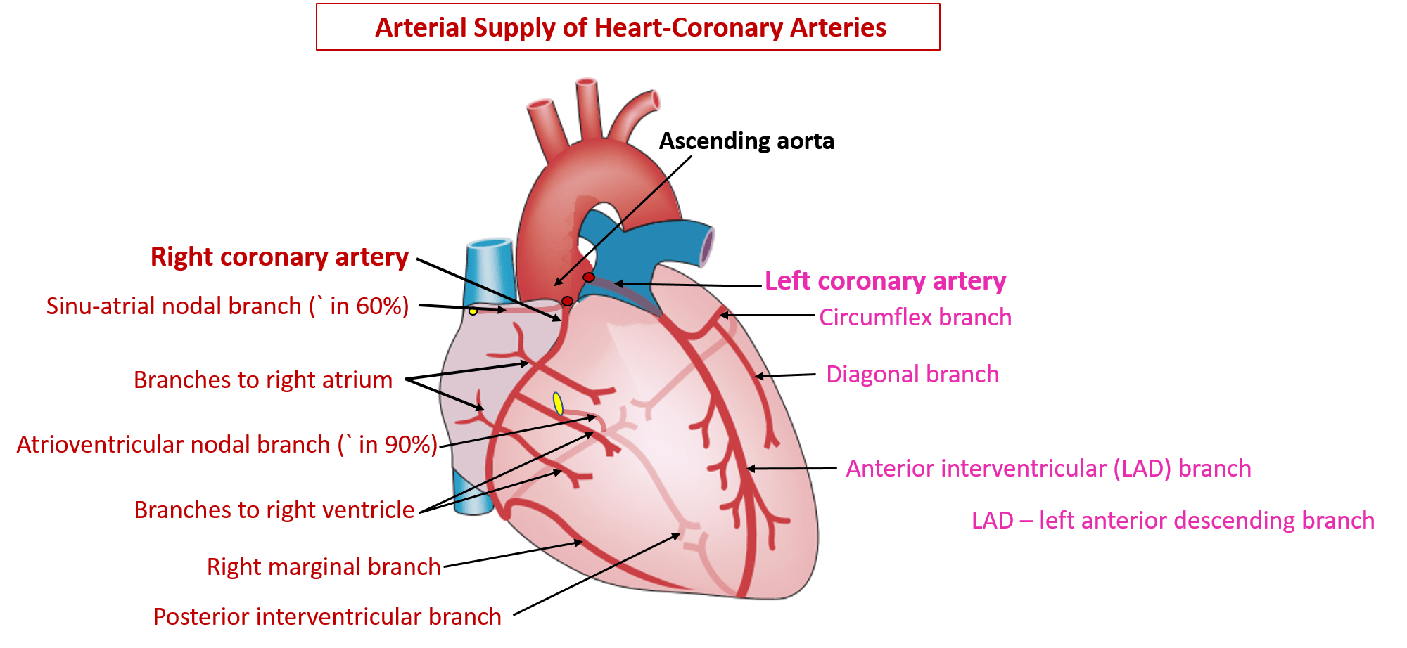
Coronary artery dominance: The artery which gives rise to the posterior interventricular artery arises determines the coronary dominance.
- If the posterior interventricular artery is a branch of right coronary artery then the coronary circulation is said to be right-dominant.
- If the posterior interventricular artery arises from circumflex artery, a branch of the left artery, then the coronary circulation is left-dominant.
- If the posterior interventricular artery arises from both the right coronary artery and the circumflex artery, then the coronary circulation is co-dominant/balanced.
Approximately 70% of the general population are right-dominant, 20% are co-dominant and 10% are left-dominant.
What Part Of The Heart Does The Posterior Descending Artery Supply
4.4/5posterior descending arterysupplies bloodheartsupplies bloodside of the heartbloodportionportionof the answer
The left coronary arteries supply:Circumflex artery – supplies blood to the left atrium, side and back of the left ventricle. Left Anterior Descending artery – supplies the front and bottom of the left ventricle and the front of the septum.
Additionally, which coronary artery supplies posterior walls? The circumflex artery branches off of the left coronary artery and supplies most of the left atrium: the posterior and lateral free walls of the left ventricle, and part of the anterior papillary muscle.
Beside above, where is the posterior descending artery located?
The posterior descending artery is also known as the posterior interventricular artery because it runs along the posterior interventricular sulcus to the apex of the heart. It is at the apex where it meets the left anterior descending artery that is traveling along the anterior surface of the heart.
Where is the left anterior descending artery?
The left anterior descending artery is the largest coronary artery runs anterior to the interventricular septum in the anterior interventricular groove, extending from the base of the heart to the apex.
Don’t Miss: Can Flonase Cause Heart Palpitations
Why Are The Coronary Arteries Important
Since coronary arteries deliver blood to the heart muscle, any coronary artery disorder or disease can have serious implications by reducing the flow of oxygen and nutrients to the heart muscle. This can lead to a heart attack and possibly death. Atherosclerosis is the most common cause of heart disease.
About Blood Supply Of Heart
The heart is a major organ of the human body which is responsible for the circulation of the blood. Blood circulation in the body ensures that every cell of the organ receives nutrients and oxygen supply to perform its metabolism. To understand the blood supply of the heart it is important to understand the location of the heart, anatomy of the heart, the vasculature of the nerve which answers questions like which artery supplies blood to the heart. This article dwells with the intricacies of blood supply of heart anatomy and functioning, which arteries supply blood to heart, the circulation system of the heart known as coronary circulation.
Also Check: How To Calculate Target Heart Rate Zone
How Does Blood Move Through The Heart
Blood comes into the right atrium from the body, moves into the right ventricle and is pushed into the pulmonary arteries in the lungs. After picking up oxygen, the blood travels back to the heart through the pulmonary veins into the left atrium, to the left ventricle and out to the bodys tissues through the aorta.
There are multiple functions going on in our bodies simultaneously. For smooth functioning and better coordination between organs, each work inside the body is differentiated so that the whole body works flawlessly.
One of the main functions of our body is the transportation of blood to and from the heart. There are two types of blood vessels arteries and veins in the circulatory system involved in carrying blood to the heart and away from the heart.
Arteries and veins, though they belong to the circulatory system, differs in their functionality and speciality. In this article, we will be looking at what exactly arteries and veins are and what are their significant differences.
Recommended Reading: Is Your Pulse And Heart Rate The Same Thing
What Are Collateral Coronary Arteries

The coronary arteries also include the collateral coronary arteries, small blood vessels that connect the normal coronary arteries with one another. When the heart is healthy, these vessels play only a minor role. When a coronary artery becomes obstructed, though, collateral coronary arteries help increase the flow of blood to the area of the heart that is being deprived of blood flow. These vessels, although small, may actually succeed in providing sufficient blood to help prevent major damage to the heart muscle during a heart attack.
Read Also: How To Calculate Target Heart Rate Zone
Overview Of Systemic Arteries
Blood relatively high in oxygen concentration is returned from the pulmonary circuit to the left atrium via the four pulmonary veins. From the left atrium, blood moves into the left ventricle, which pumps blood into the aorta. The aorta and its branchesthe systemic arteriessend blood to virtually every organ of the body.
Figure 2. The major systemic arteries shown here deliver oxygenated blood throughout the body.
Cardiac Pain Due To Angina Pectoris Or Myocardial Infarction Is Usually Referred To The Left Precordium And Medial Aspect Of Left Arm And Forearm
The heart is innervated by upper four thoracic spinal segments . The skin over the precordium is supplied by T2-T4 spinal segments and the skin over the medial aspect of forearm and arm by T1 and T2 spinal segments respectively. The cardiac pain is therefore referred to the precordium and medial aspects of arm and forearm because of the same spinal segmental innervation.
Recommended Reading: Top Part Of Heart Not Working
What Is The Name Of The Blood Vessels That Carry Blood To And From The Lungs
pulmonary arterypulmonary
Refering to the common carotid artery supplies blood to the head and face. The blood vessel that carries blood from the right ventricle to the lungs. It is the only artery that carries deoxygenated blood.
Also, what is the blood vessel that carries blood to the heart called? blood vessels: Blood moves through many tubes called arteries and veins, which together are called blood vessels. The blood vessels that carry blood away from the heart are called arteries. The ones that carry blood back to the heart are called veins.
People also ask, what carries blood away from the lungs?
Key terms
Arteries Serving The Lower Limbs
The external iliac artery exits the body cavity and enters the femoral region of the lower leg . As it passes through the body wall, it is renamed the femoral artery. It gives off several smaller branches as well as the lateral deep femoral artery that in turn gives rise to a lateral circumflex artery. These arteries supply blood to the deep muscles of the thigh as well as ventral and lateral regions of the integument. The femoral artery also gives rise to the genicular artery, which provides blood to the region of the knee. As the femoral artery passes posterior to the knee near the popliteal fossa, it is called the popliteal artery. The popliteal artery branches into the anterior and posterior tibial arteries.
Figure 11. Major arteries serving the lower limb are shown in anterior and posterior views.
Figure 12. The flow chart summarizes the distribution of the systemic arteries from the external iliac artery into the lower limb.
Don’t Miss: Is Tylenol Bad For Your Heart
Veins Draining The Lower Limbs
The superior surface of the foot drains into the digital veins, and the inferior surface drains into the plantar veins, which flow into a complex series of anastomoses in the feet and ankles, including the dorsal venous arch and the plantar venous arch. From the dorsal venous arch, blood supply drains into the anterior and posterior tibial veins. The anterior tibial vein drains the area near the tibialis anterior muscle and combines with the posterior tibial vein and the fibular vein to form the popliteal vein. The posterior tibial vein drains the posterior surface of the tibia and joins the popliteal vein. The fibular vein drains the muscles and integument in proximity to the fibula and also joins the popliteal vein. The small saphenous vein located on the lateral surface of the leg drains blood from the superficial regions of the lower leg and foot, and flows into to the popliteal vein. As the popliteal vein passes behind the knee in the popliteal region, it becomes the femoral vein. It is palpable in patients without excessive adipose tissue.
Figure 19. Anterior and posterior views show the major veins that drain the lower limb into the inferior vena cava.
Figure 20 is a flow chart of veins flowing into the lower limb. Table 14 summarizes the major veins of the lower limbs.
Figure 20. The flow chart summarizes venous flow from the lower limb.
Diagnosis And Treatment Of Coronary Artery Disease
Fig 1.6 A coronary angiogram. Two critical narrowings have been labelled.
A blockage in a coronary artery can be rapidly identified by performing a coronary angiogram. The imaging modality involves the insertion of a catheter into the aorta via the femoral artery. A contrast dye is injected into the coronary arteries and x-ray based imaging is then used to visualise the coronary arteries and any blockage that may be present.
Immediate treatment of a blockage can be performed by way of a coronary angioplasty, which involves the inflation of a balloon within the affected artery. The balloon pushes aside the atherosclerotic plaque and restores the blood flow to the myocardium. The artery may then be supported by the addition of an intravascular stent to maintain its volume.
Don’t Miss: What Heart Chamber Pushes Blood Through The Aortic Semilunar Valve
Arteries Of Lower Limbs
The artery that supplies the greater part of the lower extremity is the direct continuation of the external iliac. It runs as a single trunk from the inguinal ligament to the lower border of the popliteal fossa, where it divides into 2 branches, the anterior and posterior tibial arteries. The upper part of the main trunk is named the femoral artery, the lower part the popliteal artery.
Femoral artery
The femoral artery begins immediately behind the inguinal ligament and ends at the junction of the middle and lower thirds of the thigh, where it passes through an opening in the adductor magnus to become the popliteal artery. In the upper third of the thigh, the femoral artery is contained in the femoral triangle in the middle third of the thigh, it is contained in the adductor canal . The branches of the femoral artery are the superficial epigastric , superficial iliac circumflex, superficial external pudendal, highest genicular, deep external pudendal, muscular, and profunda femoris arteries.
Profunda femoris artery
The profunda femoris is a large vessel arising from the lateral and back part of the femoral artery that gives off the following branches: lateral femoral circumflex, medial femoral circumflex, perforating, and muscular. The terminal part of the profunda is sometimes named the fourth perforating artery.
Popliteal artery
Anterior tibial artery
Dorsalis pedis
Posterior tibial artery
Fibular artery
Which Artery Supplies Blood To The Liver

liverblood supplyhepatic arterybloodhepaticblood
. Also asked, what arteries supply the liver?
The liver is connected to two large blood vessels, the hepatic artery and the portal vein. The hepatic artery carries blood from the aorta to the liver, whereas the portal vein carries blood containing the digested nutrients from the entire gastrointestinal tract, and also from the spleen and pancreas to the liver.
One may also ask, what is unique about the blood supply to the liver? Blood flow to the liver is unique in that it receives both oxygenated and deoxygenated blood. As a result, the partial gas pressure of oxygen and perfusion pressure of portal blood are lower than in other organs of the body.
Likewise, people ask, where does the liver get its blood from?
Blood leaves the liver through the hepatic veins. This blood is a mixture of blood from the hepatic artery and from the portal vein. The hepatic veins carry blood to the inferior vena cavathe largest vein in the bodywhich then carries blood from the abdomen and lower parts of the body to the right side of the heart.
What artery carries oxygen to the liver?
Blood enters the liver through the hepatic artery and the portal vein. The blood from the hepatic artery carries oxygen and helps support liver growth. The portal vein carries blood and nutrients from the intestine and delivers them to the liver cells , which perform specific liver functions.
You May Like: Can Prednisone Cause Heart Palpitations
