Exercise During The First Trimester
If exercising is challenging during this time, or youre unsure where to begin, I always encourage first-trimester women to master the basics. In this case, the basics can involve learning how to breathe efficiently through whats termed 360-degree breathing.
This diaphragmatic breathing pattern involves inhaling and exhaling through your front, back, and sides instead of belly-breathing or shallow breathing through your neck and shoulders.
As pregnancy progresses, abdominal muscles stretch, and many women complain of a tight upper back and neck. It is vital to avoid a tight neck and shoulders by learning to create back-body expansion by breathing into the thoracic .
How Does Blood Go Back To The Heart
Blood Flow Through the Heart
blood returnsheartblood back to the heartblood
Similarly, you may ask, what body systems help blood return to the heart?
The circulatory system is made up of blood vessels that carry blood away from and towards the heart. Arteries carry blood away from the heart and veins carry blood back to the heart. The circulatory system carries oxygen, nutrients, and hormones to cells, and removes waste products, like carbon dioxide.
Beside above, what carries blood back to the heart? The blood vessels that carry blood away from the heart are called arteries. The ones that carry blood back to the heart are called veins.
Correspondingly, how long does it take a drop of blood to travel away from and back to the heart again?
Answer: On average, it takes about 45 seconds for blood to circulate from the heart, all around the body, and back to the heart again. An average adult’s heart beats more than 100,000 times a day.
How does blood flow through the body step by step?
Blood flows through your heart and lungs in four steps:
You May Like Also
What The Heart And Circulatory System Do
The circulatory system works closely with other systems in our bodies. It supplies oxygen and nutrients to our bodies by working with the respiratory system. At the same time, the circulatory system helps carry waste and carbon dioxide out of the body.
Hormones produced by the endocrine system are also transported through the blood in the circulatory system. As the bodys chemical messengers, hormones transfer information and instructions from one set of cells to another. For example, one of the hormones produced by the heart helps control the kidneys release of salt from the body.
One complete heartbeat makes up a cardiac cycle, which consists of two phases:
In the systemic circulation, blood travels out of the left ventricle, to the aorta, to every organ and tissue in the body, and then back to the right atrium. The arteries, capillaries, and veins of the systemic circulatory system are the channels through which this long journey takes place.
Don’t Miss: Fluticasone Heart Palpitations
What Is Vascular Disease
A vascular disease is a condition that affects the arteries and veins. Most often, vascular disease affects blood flow, either by blocking or weakening blood vessels, or by damaging the valves that are found in veins. Organs and other body structures may be damaged by vascular disease as a result of decreased or completely blocked blood flow.
Pregnancy Exercises During Third Trimester

As the bodys center of mass changes, it becomes increasingly essential to err on the side of caution when it comes to single-leg or balancing exercises. Performing Bulgarian split squats or single-leg deadlifts close to a wall will increase stability and reduce the risk of falling.
The growing belly can also exacerbate postural changes. Many third-trimester women welcome mobility exercises that provide relief for the lower back, particularly if they tend to stand with an anterior pelvic tilt .
To help with this, perform pelvic tucks/tilts by lying on the floor with bent knees and practice pulling your belly button in toward your spine while pushing your pelvis up towards the ceiling. Rotating between a neutral pelvis and a posterior pelvic tilt can help mobilize the hip and release lower back tension.
Read Also: What Should Your Resting Heart Beat Be
Which Blood Vessels Carry Deoxygenated Blood Away From The Heart
pulmonary arteries
. Likewise, people ask, which blood vessels carry blood away from the heart?
The arteries carry oxygen and nutrients away from your heart, to your body’s tissues. The veins take oxygen-poor blood back to the heart. Arteries begin with the aorta, the large artery leaving the heart. They carry oxygen-rich blood away from the heart to all of the body’s tissues.
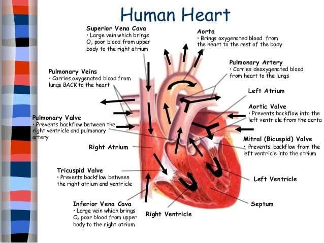
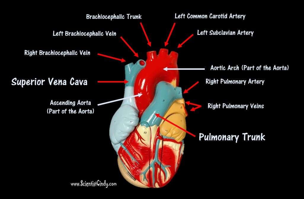
what vessels carry deoxygenated blood to the right atrium? Great VesselsThe superior vena cava and the inferior vena cava bring deoxygenated blood from the body to the right atrium. The pulmonary trunk exits from the right ventricle and carries deoxygenated blood to the lungs.
People also ask, which of the following blood vessels carries deoxygenated blood?
The heart
| Carries deoxygenated blood from the body back to the heart. | |
| Pulmonary artery | Carries deoxygenated blood from the heart to the lungs. |
| Pulmonary vein | Carries oxygenated blood from the lungs to the heart. |
| Aorta | Carries oxygenated blood from the heart around the body. |
Do veins carry blood away from the heart?
Most veins carry deoxygenated blood from the tissues back to the heart exceptions are the pulmonary and umbilical veins, both of which carry oxygenated blood to the heart. In contrast to veins, arteries carry blood away from the heart.
What Part Of The Heart Pumps Blood To The Rest Of The Body
The right ventricle pumps the oxygen-poor blood to the lungs through the pulmonary valve. The left atrium receives oxygen-rich blood from the lungs and pumps it to the left ventricle through the mitral valve. The left ventricle pumps the oxygen-rich blood through the aortic valve out to the rest of the body.
Recommended Reading: Carrie Fisher Brain Damage
Valves Maintain Direction Of Blood Flow
As the heart pumps blood, a series of valves open and close tightly. These valves ensure that blood flows in only one direction, preventing backflow.
- The tricuspid valve is situated between the right atrium and right ventricle.
- The pulmonary valve is between the right ventricle and the pulmonary artery.
- The mitral valve is between the left atrium and left ventricle.
- The aortic valve is between the left ventricle and the aorta.
Each heart valve, except for the mitral valve, has three flaps that open and close like gates on a fence. The mitral valve has two valve leaflets.
How Blood Flows Through The Heart And Lungs
The heart is a complex organ, using four chambers, four valves, and multiple blood vessels to provide blood to the body. Blood flow itself is equally complex, involving a cyclic series of steps that move blood trough the heart and to the lungs to be oxygenated, deliver it throughout the body, then bring blood back to the heart to re-start the process.
This is the key function of the cardiovascular system: consuming, transporting, and using oxygen throughout physical activity . Disruptions in blood flow through the heart and lungs can have serious effects.
Don’t Miss: How To Find Thrz
Which Blood Vessel Takes Oxygen Rich Blood From The Lungs And Brings It Back To The Heart
pulmonary arterypulmonary veins
. Similarly, which blood vessel takes blood from the lungs to the heart?
pulmonary artery: A blood vessel that carries blood from the heart to the lungs, where the blood picks up oxygen and then returns to the heart. pulmonary vein: One of four veins that carry oxygen-rich blood from the lungs to the heart.
Subsequently, question is, what part of the heart receives oxygen rich blood from the lungs? The right ventricle pumps the oxygen–poor blood to the lungs through the pulmonary valve. The left atrium receives oxygen–rich blood from the lungs and pumps it to the left ventricle through the mitral valve. The left ventricle pumps the oxygen–rich blood through the aortic valve out to the rest of the body.
Subsequently, question is, which blood vessel sends oxygen rich blood from the heart to the rest of the body?
The arteries carry oxygen and nutrients away from your heart, to your body’s tissues. The veins take oxygen-poor blood back to the heart. Arteries begin with the aorta, the large artery leaving the heart. They carry oxygen-rich blood away from the heart to all of the body’s tissues.
How many vessels return oxygenated blood to the heart?
The oxygenated blood from the lungs now returns to the left atrium via four tubes that are known as pulmonary veins . The pulmonary veins empty into the back portion of the LA.
Whats The Difference Between Veins And Arteries
verifiedMelissa Petruzzello
Veins and arteries are major players in the circulatory system of all vertebrates. They work together to transport blood throughout the body, helping to oxygenate and remove waste from every cell with each heartbeat. Arteries carry oxygenated blood from the heart, while veins carry oxygen-depleted blood back to the heart. An easy mnemonic is “A for artery and away .”
As the vessels that are closest to the heart, arteries must contend with intense physical pressure from the blood moving forcibly through them. They pulse with each heartbeat and have thicker walls. Veins experience much less pressure but must contend with the forces of gravity to get blood from the extremities back to the heart. Many veins, especially those in the legs, have valves to prevent the backflow and pooling of blood. Although veins are often depicted as blue in medical diagrams and sometimes appear blue through pale skin, they are not actually blue in color. Light interacts with skin and deoxygenated blood, which is a darker shade of red, to reflect a blue tone. Veins seen during surgery or in cadavers look nearly identical to arteries.
Recommended Reading: Low Pulse High Blood Pressure Causes
What Are The Different Types Of Arteries
There are three types of arteries. Each type is composed of three coats: outer, middle, and inner.
- Elastic arteries are also called conducting arteries or conduit arteries. They have a thick middle layer so they can stretch in response to each pulse of the heart.
- Muscular arteries are medium-sized. They draw blood from elastic arteries and branch into resistance vessels. These vessels include small arteries and arterioles.
- Arterioles are the smallest division of arteries that transport blood away from the heart. They direct blood into the capillary networks.
There are four types of veins:
- Deep veins are located within muscle tissue. They have a corresponding artery nearby.
- Superficial veins are closer to the skins surface. They dont have corresponding arteries.
- Pulmonary veins transport blood thats been filled with oxygen by the lungs to the heart. Each lung has two sets of pulmonary veins, a right and left one.
- Systemic veins are located throughout the body from the legs up to the neck, including the arms and trunk. They transport deoxygenated blood back to the heart.
Use this interactive 3-D diagram to explore an artery.
Use this interactive 3-D diagram to explore a vein.
If The Saphenous Vein Is Removed How Is The Body Able To Circulate Blood

Small veins give deoxigenated blood to the main saphenous vein. After that, deoxygenated blood goes through the saphenous to the heart and lungs to get fresh oxygen and circulate it through arteries. If you remove the saphenous, how does this process work? How is the body able to circulate blood without it?
A healthy saphenous vein assists in returning blood from the lower extremities to the heart, but this function is dependent on the presence of a healthy system of check valves throughout the vein.In the presence of venous insufficiency, where the valves are not working, blood runs in the wrong direction from the body down to the lower leg and actually overloads the other veins that are trying to return blood to the heart.Under normal circumstances, 70-80% of the blood returning to the heart comes through the deep veins in the muscles of the leg. In the circumstances of saphenous vein reflux or varicose veins, these deep veins have to work extra hard to return the blood to the heart. After removal or ablation of a diseased saphenous vein, the blood will flow through the deep system in a normal manner. In most instances, the deep system has more than enough excess capacity to handle the blood that would normally go through the saphenous vein.
The saphenous vein is not a major vein and, in fact, provides only a tiny amount of venous return. It is a superficial vein and all of the superficial veins in the lower leg only account for 10% of venous return.
Read Also: What Causes Bleeding Around The Heart
Which Heart Artery Is The Widowmaker
The widow-maker is a massive heart attack that occurs when the left anterior descending artery is totally or almost completely blocked. The critical blockage in the artery stops, usually a blood clot, stops all the blood flow to the left side of the heart, causing the heart to stop beating normally.
Vasculature Of The Head
Blood is carried from your heart to the rest of your body through a complex network of arteries, arterioles, and capillaries. Blood is returned to your heart through venules and veins.
The one-way vascular system carries blood to all parts of your body. This process of blood flow within your body is called circulation. Arteries carry oxygen-rich blood away from your heart, and veins carry oxygen-poor blood back to your heart.
In pulmonary circulation, though, the roles are switched. It is the pulmonary artery that brings oxygen-poor blood into your lungs and the pulmonary vein that brings oxygen-rich blood back to your heart.
Like the heart, the brains cells need a constant supply of oxygen-rich blood. This blood supply is delivered to the brain by the two large carotid arteries in the front of your neck and by two smaller vertebral arteries at the back of your neck.
The right and left vertebral arteries come together at the base of your brain to form what is called the basilar artery in the vasculature of your head.
As shown in the diagrams of the vasculature of the head, the vessels that carry oxygen-rich blood are colored red, and the vessels that carry oxygen-poor blood are colored blue.
You May Like: Does Tylenol Increase Heart Rate
How Does Blood Flow Through The Heart
The right and left sides of the heart work together. The pattern described below is repeated over and over, causing blood to flow continuously to the heart, lungs, and body.
Right side of the heart
- Blood enters the heart through two large veins, the inferior and superior vena cava, emptying oxygen-poor blood from the body into the right atrium.
- As the atrium contracts, blood flows from your right atrium into your right ventricle through the open tricuspid valve.
- When the ventricle is full, the tricuspid valve shuts. This prevents blood from flowing backward into the right atrium while the ventricle contracts.
- As the ventricle contracts, blood leaves the heart through the pulmonic valve, into the pulmonary artery and to the lungs, where it is oxygenated. The oxygenated blood then returns to the heart through the pulmonary veins.
Left side of the heart
- The pulmonary veins empty oxygen-rich blood from the lungs into the left atrium.
- As the atrium contracts, blood flows from your left atrium into your left ventricle through the open mitral valve.
- When the ventricle is full, the mitral valve shuts. This prevents blood from flowing backward into the atrium while the ventricle contracts.
- As the ventricle contracts, blood leaves the heart through the aortic valve, into the aorta and to the body.
Electrical Impulses Keep The Beat
The heart’s four chambers pump in an organized manner with the help of electrical impulses that originate in the sinoatrial node . Situated on the wall of the right atrium, this small cluster of specialized cells is the heart’s natural pacemaker, initiating electrical impulses at a normal rate.
The impulse spreads through the walls of the right and left atria, causing them to contract, forcing blood into the ventricles. The impulse then reaches the atrioventricular node, which acts as an electrical bridge for impulses to travel from the atria to the ventricles. From there, a pathway of fibers carries the impulse into the ventricles, which contract and force blood out of the heart.
Read Also: Does Tylenol Increase Heart Rate
What Makes The Heart Pump
Your heart has a special electrical system called the cardiac conduction system. This system controls the rate and rhythm of the heartbeat. With each heartbeat, an electrical signal travels from the top of the heart to the bottom. As the signal travels, it causes the heart to contract and pump blood.
Also Check: What Heart Chamber Pushes Blood Through The Aortic Semilunar Valve
Classification & Structure Of Blood Vessels
Blood vessels are the channels or conduits through which blood is distributed to body tissues. The vessels make up two closed systems of tubes that begin and end at the heart. One system, the pulmonary vessels, transports blood from the right ventricle to the lungs and back to the left atrium. The other system, the systemic vessels, carries blood from the left ventricle to the tissues in all parts of the body and then returns the blood to the right atrium. Based on their structure and function, blood vessels are classified as either arteries, capillaries, or veins.
You May Like: Can Ibs Cause Heart Palpitations
You May Like: Gerd Tachycardia
