What Causes Silent Heart Attacks
Silent heart attacks are caused by the same things that cause traditional heart attacks. This happens when part of the heart muscle is damaged or dies because it hasnt received enough oxygen. This is often due to a blocked artery in the heart. Risk factors for silent heart attacks are also the same. They include:
- Smoking.
- Gender women have silent heart attacks more often than men.
Why Is It Done
An ECG gives two major kinds of information. First, by measuring time intervals on the ECG, a doctor can determine how long the electrical wave takes to pass through the heart. Finding out how long a wave takes to travel from one part of the heart to the next shows if the electrical activity is normal or slow, fast or irregular. Second, by measuring the amount of electrical activity passing through the heart muscle, a cardiologist may be able to find out if parts of the heart are too large or are overworked.
Can Wearables Detect A Heart Attack
Last, were often asked, Can Apple Watch detect a heart attack? Its a natural question, but there are a few of things to remember.
First off, a heart attack is only one of three lethal outcomes in heart disease the other two, stroke and cardiac arrest, kill more people. Second, heart attack survival rate is currently about 95%, compared with 60% for stroke and 8% for cardiac arrest, so the potential benefit of detection is higher for the latter two. Third, if youre having a heart attack, your heart rate may be lower, higher, or the same as average, which makes it hard to diagnose heart attacks by an optical sensor. The tests people use today are the ECG and troponin.
Those factors together suggest heart rhythm are the more likely targets for current wearable technology. Of course, future Apple Watches may include an ECG sensor, in which case even more possibilities will emerge.
Recommended Reading: Does Tylenol Increase Heart Rate
How Is A Heart Attack Diagnosed
As soon as ambulance or medical staff arrive they will begin tests to find out what is happening to you. These will include:
- An ECG : to show the amount of damage to your heart muscle and where the damage is. Treatment to restore blood flow and minimise the amount of heart muscle damage can be achieved in different ways depending on your ECG readings so it is important that you have an ECG as soon as possible to show exactly what is happening.
- Blood tests: the main blood test is to measure the amount of troponin in your blood. Troponin is a protein that is released into your blood stream when your heart muscle is damaged, for example during a heart attack. The level of troponin in your blood is increased if you have had a heart attack.
Living With Silent Heart Attacks

After youve had a silent heart attack, you are at higher risk of having another one. This one will likely be more severe and harmful. Your doctor will likely recommend heart-healthy lifestyle changes to help reduce your risk. They include:
- A heart-healthy diet.
- Working toward a healthy weight.
- Managing stress.
- Being physically active.
- Quitting smoking.
Symptoms during a second heart attack may be different than the first one. If you have any new symptoms of a heart attack or are in any doubt, call 911. Early treatment is the key to surviving a heart attack.
Don’t Miss: Reflux And Palpitations
What Do Normal And Abnormal Heart Rhythms Look Like On Apple Watch
Almost every month, a news storypops up about somebody whoselife was saved by their Apple Watch. As part of the mRhythm Study, were analyzing a lot of heart rate data, and decided to write a brief primer on what both normal and abnormal heart rhythms look like when measured on an Apple Watch.
Download Cardiogram for your Apple Watch, Fitbit, wearOS, or Garmin watch here:
Note: although we believe wearables can save livesand hope to play a part in making that happenthere is a lot of clinical groundwork that needs to happen first. Please dont take any of the below as medical advice.
Heart Attack Ekg Evidence
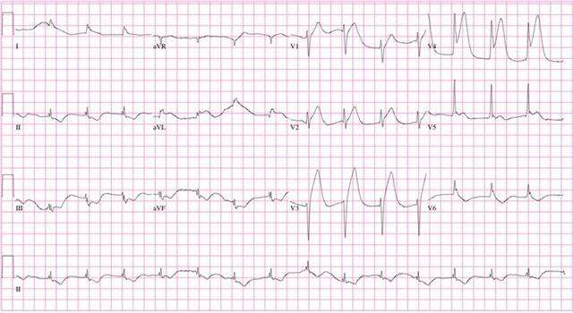
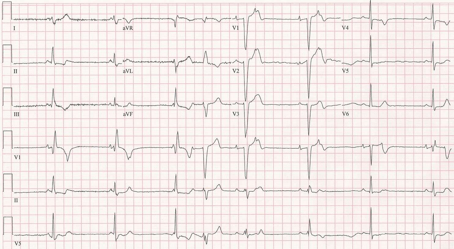
The two clues that make it possible to identify a heart attack by type are ischemic EKG changes in the ST segment and the appearance of Q waves. The ST segment may become elevated, depressed, or may not change at all during a heart attack. If a change does occur it usually does not persist unless an aneurysm of the heart muscle forms, in which case it adjoins a Q wave. Q waves usually, but not always, form later in the process of the acute event or after it is complete.
Q waves might appear in leads where they were not prior to the heart attack or in place of R waves. When Q waves replace R waves the initial deflection representing contraction of the ventricles is downward rather than upward. Either type of change can appear in leads whose sensors face the damaged area of the heart.
Don’t Miss: Can Ibs Cause Heart Palpitations
How The Test Is Performed
You will be asked to lie down. The health care provider will clean several areas on your arms, legs, and chest, and then will attach small patches called electrodes to those areas. It may be necessary to shave or clip some hair so the patches stick to the skin. The number of patches used may vary.
The patches are connected by wires to a machine that turns the heart’s electrical signals into wavy lines, which are often printed on paper. The doctor reviews the test results.
You will need to remain still during the procedure. The provider may also ask you to hold your breath for a few seconds as the test is being done.
It is important to be relaxed and warm during an ECG recording because any movement, including shivering, can alter the results.
Sometimes this test is done while you are exercising or under light stress to look for changes in the heart. This type of ECG is often called a stress test.
How To Prepare For An Ekg
You normally dont need to follow any special instructions in order to prepare for an EKG. Your doctor will want to know about medications you take to make sure that they wont interfere with the results of your EKG. You dont need to fast or make any dietary changes before having this type of test done.
Dont Miss: Can Acid Reflux Cause Heart Palpitations
Also Check: What Causes Bleeding Around The Heart
What Is A Heart Attack: What Does An Ekg Look Like In A Heart Attack
A heart attack occurs when one of the arteries in the heart is occluded or semi included so that the blood and oxygen cannot get to the muscle.
There are many reasons that a heart attack can happen. These EKG Calipers are used to measure exact measurements on an EKG. This book called The Rapid Interpretation of EKGs, has illustrations on each page on how to interpret EKGs.
The following are risks for heart attack :
- Coronary artery disease
- Inactivity
According to the Centers for Disease Control, one million American have heart attacks each year. Around 250,000 of thee people have previously had a heart attack. Almost 20% of people who have had a first heart attack will die.
What Does an EKG Look Like In a Heart Attack With pictures? How does an old heart attack look like in a heart attack?
Here’s A Rundown Of The Likely Chain Of Events After You Call 911
For a suspected heart attack, paramedics can often perform initial testing in the ambulance while the patient is en route to the hospital. Image: Thinkstock
Every 43 seconds, someone in the United States has a heart attack. If one day that someone is you or a loved one, it may be helpful to know what’s likely to happen, both en route to the hospital and after you arrive.
For starters, always call 911 to be transported via ambulance rather than going by car. Contrary to what you might assume, speed isn’t the only rationale. “If you’re having a heart attack, there are two reasons why you want to be in an ambulance,” says Dr. Joshua Kosowsky, assistant professor of emergency medicine at Harvard Medical School. One is that in the unlikely event of cardiac arrest, the ambulance has the equipment and trained personnel to restart your heart. Cardiac arrest, which results from an electrical malfunction that stops the heart’s pumping ability, is fatal without prompt treatment. However, most heart attacks do not cause cardiac arrest, Dr. Kosowsky stresses. “It’s rare, but it’s certainly not a risk you want to take while you’re driving or riding in a car.”
Read Also: Can Flonase Cause Heart Palpitations
Silent Heart Attack Treatment
Normally, silent heart attacks are found long after the heart attack is over. Treatment will mostly involve taking medicines. These medicines help improve blood flow to your heart, prevent clotting, and reduce the risks of having another heart attack. They include:
- aspirin
- ACE inhibitors
- fish oil
Your doctor will prescribe the medicines that are right for you. If you have had a heart attack, your doctor will also talk to you about lifestyle changes. You can make these changes to prevent more heart problems.
Stemi Vs Nstemi Which Is Worse

The bottom line is that both are just as bad. STEMI is seen as more of an immediate emergency because there is a known total occlusion of a heart vessel that needs opening back up urgently. In terms of long-term outcomes, they have equal health implications. Patients with NSTEMI often have other illnesses such as ongoing critical illness, diabetes, kidney disease, and other that means they have a generally high risk over the long term. Both STEMI and NSTEMI need aggressive treatment over the short and long term.
Read Also: Thrz Calculator
What Does A Normal Ekg Look Like Versus An Abnormal One From An Undetected Heart Attack
Ask U.S. doctors your own question and get educational, text answers â it’s anonymous and free!
Ask U.S. doctors your own question and get educational, text answers â it’s anonymous and free!
HealthTap doctors are based in the U.S., board certified, and available by text or video.
Blood Tests And Beyond
But not all heart attacks show up on the first ECG. So even if it looks normal, you’re still not out of the woods, says Dr. Kosowsky. The next step is an evaluation by a doctor or other clinician, who will ask about your medical history and details about the location, duration, and intensity of your symptoms. You’ll also have a blood test to measure troponin, a protein that rises in response to heart muscle damage. This blood test is very sensitive. But keep in mind that elevated levels don’t always show up right away. That’s why doctors sometimes have people stay for several hours to get a follow-up troponin measurement.
Other possible tests include a chest x-ray to look for alternative causes of chest discomfort, such as pneumonia or heart failure. A doctor also might give you a trial of medication to see whether it relieves your symptoms, and additional ECGs may be performed over time.
Often, if several troponin tests come back normal, the doctor may want to check your risk of a future heart attack with an exercise stress test. This test can reveal how your heart responds to the demands of increased blood flow needed during exercise. During a standard exercise test, you walk on a treadmill at progressively faster speeds, while trained staff monitors your heart’s electrical activity, your heart rate, and your blood pressure.
Recommended Reading: Ibs And Palpitations
Type #: Posterior St Segment Elevation Mi
This one is tricky when isolated, but it is very important not to miss. We treat it just like any other ST segment elevation MI, which is of course time sensitive.
The posterior wall is supplied by the posterior descending artery. The PDA branches from the right coronary artery in 80% of people therefore, occlusion of RCA can result in both an inferior STEMI and a posterior MI as well. Sometimes, it is obvious on the ECG when a posterior MI accompanies an inferior STEMI, but it can also occur all by itself.
The ECG criteria to diagnose a posterior MI treated like a STEMI, even though no real ST segment elevation is apparent include:
- ST segment depression in V1 to V4. Think of things backwards. These are the septal and anterior ECG leads. The MI is posterior , so there is ST depression instead of elevation. Turn the ECG upside down, and it would look like a STEMI.
- The ratio of the R wave to the S wave in leads V1 or V2 is greater than 1. This represents an upside-down Q wave .
- ST segment elevation in the posterior leads of a posterior ECG . A posterior ECG is done by simply adding three extra precordial leads wrapping around the left chest wall toward the back.
Below are some examples including isolated posterior MIs, inferior STEMIs with posterior involvement and a posterior ECG.
Below are two examples of ECG tracings with both inferior STEMI and posterior involvement. Remember, the more you look at the better!
Inferior-Posterior STEMI Example #1
Key Background: Ecg Vs Ppg
Before explaining the details of the abnormal rhythms, it helps to have a little background on the two most common types of heart rate sensors.
The gold standard in a hospital setting is an electrocardiogram, or ECG, which shows the voltage within your hearts electrical system. In every normal heart beat, the top two chambers of the heart contract first, producing a small voltage notch called the P wave. Specialized electrical tissue called the AV node causes a 120200ms delay, and then the bottom two chambers of the heart contract, producing a larger voltage spike called the QRS complex. In normal heart rhythm, every QRS complex follows a P wave .
A cardiologist can infer quite a lot from even a short ECG reading. For example, if the QRS complex precedes or obscures the P wave, it indicates electrical impulses in the heart are traveling in reverse , which can happen in a common arrhythmia called supraventricular tachycardia .
Consumer heart rate sensors, such as the Apple Watch, use an optical sensor based on photoplethysmography . Every time your heart beats, it sends a pulse wave throughout your arteries. Apple Watch measures those pulse waves at the wrist, but anywhere on the skin with good blood perfusione.g., your forehead, arm, chest, ear lobescan be used to measure heart rate.
Read Also: Does Tylenol Increase Heart Rate
Type #: Anterior St Segment Elevation Mi
This is the big one that carries a high mortality if not treated rapidly. An anterior STEMI is usually from acute thrombotic occlusion of the left anterior descending coronary artery also known as the widow maker.
Recall that the anterior leads are technically V3 and V4 however, it is common for the septum and/or lateral wall to be involved during anterior MIs, as the LAD supplies septal branches to the interventricular septum and diagonal branches to the lateral wall. The septum is represented on the ECG by leads V1 and V2, whereas the lateral wall is represented by leads V5, V6, lead I and lead aVL. To make things more complicated, sometimes the LAD wraps around the cardiac apex, which is a common anatomic variant. This results in part of the inferior wall being supplied by the LAD, as well. Oh, my!
We will get to the examples soon, but first we need to understand some more basics of anterior MIs.
If the thrombus is in the proximal LAD, the septum and lateral walls will often also be involved, in addition to the anterior segments, resulting in ST segment elevation in leads V1 through V6 and perhaps lead I and aVL, as well.
When the thrombus is in the mid LAD , the diagonal branch may or may not be involved. If this is the case, then the ST segment elevation will be in V3 to V6 and not the septal leads.
Below are the anterior MI ECG patterns that you may encounter.
Anterior MI Pattern Tombstoning
Anterior MI Pattern Typical ST Segment Elevation
About Half Of All Heart Attacks Are Mistaken For Less Serious Problems And Can Increase Your Risk Of Dying From Coronary Artery Disease
You can have a heart attack and not even know it. A silent heart attack, known as a silent myocardial infarction , account for 45% of heart attacks and strike men more than women.
They are described as “silent” because when they occur, their symptoms lack the intensity of a classic heart attack, such as extreme and pressure stabbing pain in the arm, neck, or jaw sudden shortness of breath sweating, and dizziness.
“SMI symptoms can feel so mild, and be so brief, they often get confused for regular discomfort or another less serious problem, and thus men ignore them,” says Dr. Jorge Plutzky, director of the vascular disease prevention program at Harvard-affiliated Brigham and Women’s Hospital.
For instance, men may feel fatigue or physical discomfort and chalk it up to overwork, poor sleep, or some general age-related ache or pain. Other typical symptoms like mild pain in the throat or chest can be confused with gastric reflux, indigestion, and .
Also, the location of pain is sometimes misunderstood. With SMI, you may feel discomfort in the center of the chest and not a sharp pain on the left side of the chest, which many people associate with a heart attack. “People can even feel completely normal during an SMI and afterward, too, which further adds to the chance of missing the warning signs,” says Dr. Plutzky.
Don’t Miss: Does Benadryl Lower Heart Rate
