Respiratory Adaptation To High Altitude
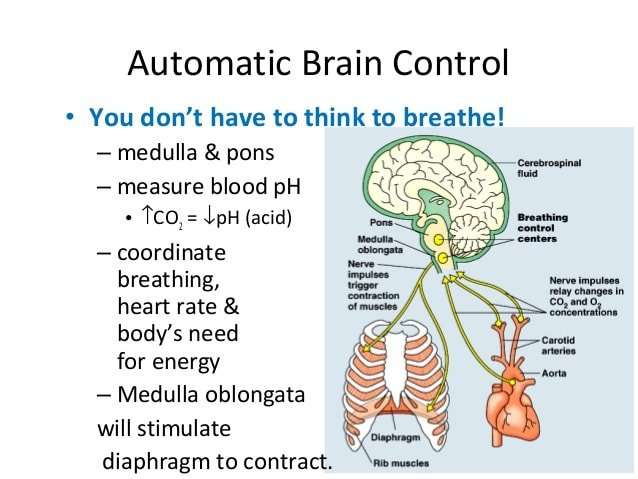
Any fall in the partial pressure of oxygen in blood is quickly detected by the peripheral chemoreceptors located in the carotid bodies. In response, they signal to the respiratory center located in the medulla oblongata of the brainstem to increase ventilation. This process is known as the hypoxic ventilatory response and its magnitude varies widely between individuals. Those with a brisk HVR show a large increase in minute volume compared to those with a blunted HVR when exposed to same degree of hypoxemia. Hyperventilation initiated by the HVR removes alveolar carbon dioxide more rapidly and thus creates a higher alveolar partial pressure of oxygen according to the alveolar gas equation.
The simplified alveolar gas equation:
PAO2 = alveolar partial pressure of oxygen Patm = atmospheric pressure PH2O = the saturated vapor pressure of water PaCO2 = arterial partial pressure of carbon dioxide RQ = respiratory quotient.
Donald Simon Urquhart, Florian Gahleitner, in, 2022
Structure Of The Medulla Oblongata
The region between the anterior median and anterolateral sulci is occupied by an elevation on either side known as the pyramid of medulla oblongata. This elevation is caused by the corticospinal tract. In the lower part of the medulla, some of these fibers cross each other, thus obliterating the anterior median fissure. This is known as the decussation of the pyramids. Other fibers that originate from the anterior median fissure above the decussation of the pyramids and run laterally across the surface of the pons are known as the external arcuate fibers.
The region between the anterolateral and posterolateral sulcus in the upper part of the medulla is marked by a swelling known as the olivary body, caused by a large mass of gray matter known as the inferior olivary nucleus.
The posterior part of the medulla between the posterior median and posterolateral sulci contains tracts that enter it from the posterior funiculus of the spinal cord. These are the fasciculus gracilis, lying medially next to the midline, and the fasciculus cuneatus, lying laterally.
The lower part of the medulla, immediately lateral to the fasciculus cuneatus, is marked by another longitudinal elevation known as the tuberculum cinereum. It is caused by an underlying collection of gray matter known as the spinal nucleus of the trigeminal nerve. The gray matter of this nucleus is covered by a layer of nerve fibers that form the spinal tract of the trigeminal nerve.
What Parts Of The Brain Is Responsible For Respiration
Now that we have that covered, lets talk about the involvement of the brain in this process.
Your brain starts where the spinal cord enters the skull, and the first section that you encounter is called the Brain Stem. The brain stem contains the following structures:
- The medulla oblongata
- The Pons
- The Midbrain
The medulla oblongata is involved in regulating many of the bodily processes that are controlled automatically like blood pressure, heart rate and yes, you guessed it . . . RESPIRATION.
The way this works is relatively straightforward. The medulla oblongata basically detects carbon dioxide and Oxygen levels in the bloodstream and determines what changes need to happen in the body.
It can then send nerve impulses to muscles in the heart and diaphragm, letting them know that they need to either step up their game or slow down a bit.
The reason I mentioned the heart is because the respiratory system is very much tied to the circulatory system.
Don’t Miss: List The Steps Of How To Calculate Your Target Heart Rate Zone
How Brain Death Occurs
Brain death can occur when the blood and/or oxygen supply to the brain is stopped. This can be caused by:
- cardiac arrest when the heart stops beating and the brain is starved of oxygen
- heart attack a serious medical emergency that occurs when the blood supply to the heart is suddenly blocked
- stroke a serious medical emergency that occurs when the blood supply to the brain is blocked or interrupted
- blood clot a blockage in a blood vessel that disturbs or blocks the flow of blood around your body
Brain death can also occur as a result of:
Parasympathetic Nervous System And Your Heart

There are a number of special receptors for the PSNS in your heart called muscarinic receptors. These receptors inhibit sympathetic nervous system action. This means theyre responsible for helping you maintain your resting heart rate. For most people, the resting heart rate is between 60 and 100 beats per minute.
On the other hand, the sympathetic nervous system increases heart rate. A faster heart rate pumps more oxygen-rich blood to the brain and lungs. This can give you the energy to run from an attacker or heighten your senses in another scary situation.
According to an article in the journal Circulation from the American Heart Association, a persons resting heart rate can be one indicator of how well a persons PSNS, specifically the vagus nerve, is working. This is usually only the case when a person doesnt take medications that affect heart rate, like beta-blockers, or have medical conditions affecting the heart.
For example, heart failure reduces the response of the parasympathetic nervous system. The results can be an increased heart rate, which is the bodys way of trying to improve the amount of blood it pumps through the body.
Don’t Miss: Flonase Chest Pain
Anatomy Of The Autonomic Nervous System
The autonomic nervous system Autonomic nervous system The peripheral nervous system consists of more than 100 billion nerve cells that run throughout the body like strings, making connections with the brain, other parts of the body, and… read more is the part of the nervous system that supplies the internal organs, including the blood vessels, stomach, intestine, liver, kidneys, bladder, genitals, lungs, pupils, heart, and sweat, salivary, and digestive glands.
The autonomic nervous system has two main divisions:
-
Sympathetic
-
Parasympathetic
After the autonomic nervous system receives information about the body and external environment, it responds by stimulating body processes, usually through the sympathetic division, or inhibiting them, usually through the parasympathetic division.
An autonomic nerve pathway involves two nerve cells. One cell is located in the brain stem Brain stem The brains functions are both mysterious and remarkable, relying on billions of nerve cells and the internal communication between them. All thoughts, beliefs, memories, behaviors, and moods… read more or spinal cord. It is connected by nerve fibers to the other cell, which is located in a cluster of nerve cells . Nerve fibers from these ganglia connect with internal organs. Most of the ganglia for the sympathetic division are located just outside the spinal cord on both sides of it. The ganglia for the parasympathetic division are located near or in the organs they connect with.
Analysis Of Blood Pressure And Heart Rate Variability
As mentioned in the introduction, cardiovascular variability was evaluated by making use of the spectral analysis. Briefly, each SBP, DBP, and PI series was split into short term data records, each lasting 512 seconds, and for each record the power spectrum was estimated by the fast Fourier transform. A typical output of this procedure is illustrated in figure , panels C and D. The spectral characteristics remained quite stable in the before BD and after BD segments, thus the spectra falling in each of these segments have been averaged to obtain a single spectrum for each condition. The respiratory component was easily identified in each spectrum by visual inspection, appearing as a clear and sharp spectral peak at frequencies higher than 0.1 Hz. The magnitude of the 10 seconds rhythm was quantified by integrating the spectrum between 0.06 and 0.12 Hz. The slowest components of variability have been globally quantified by estimating the exponent of the 1/f law relating the power of heart rate or blood pressure spectra with the frequency f at the lower frequencies of the spectrum. When both the vertical and horizontal axes of a spectrum are represented in a log scale, the 1/f trend is transformed into a linear trend with slope . Thus, once the spectrum was plotted in a log-log scale, the exponent was estimated by computing the slope of the regression line between power and frequency in the band ranging from 4×10⁻³ to 2×10⁻² Hz.
You May Like: Will Tylenol Raise Blood Pressure
Analysis Of Blood Pressure And Heart Rate Variabilities
The spectra of a representative patient are shown in figure . Important changes were associated with brain death. The mechanical ventilator, set at 12 cycles/minute in this patient, produced a clear cut respiratory peak at 0.2 Hz in blood pressure and PI spectra. In the PI spectrum the amplitude of such a peak dropped by more than 99% after brain death. The amplitude of the respiratory peak remained almost unchanged in the SBP and DBP spectra after death. In this case, however, it was the power of spectral components surrounding the respiratory peak which dramatically reduced. Brain stem death also resulted in a drastic power fall at 0.1 Hz and in a steepening of the 1/f line representing the trend of the slowest components of variability in SBP, DBP, and PI spectra.
SBP, DBP, and PI power spectral densities in a representative patient before and after brain death. Because the spectra span over four decades of power and three decades of frequency, a log-log scale is used to facilitate the identification of spectral details. Abbreviations as in fig .
Mean of the slope of the regression line fitting the spectrum in a log-log scale
Squared coherence modulus between SBP and PI, k2SBP-PI, in a representative patient , before and after brain death.
What Is The Medulla Oblongata And What Does It Do
For most of the 18th century, the medulla oblongata was thought to simply be an extension of the spinal cord without any distinct functions of its own. This changed in 1806, when Julien-Jean-Cesar Legallois found that he could remove the cortex and cerebellum of rabbits and they would continue to breathe. When he removed a specific section of the medulla, however, respiration stopped immediately. Legallois had found what he believed to be a “respiratory center” in the medulla, and soon after the medulla was considered to be a center of vital functions .
Over time, exactly which “vital functions” were linked to the medulla would become more clear, and the medulla would come to be recognized as a crucial area for the control of both cardiovascular and respiratory functions. The role of the medulla in cardiovascular function involves the regulation of heart rate and blood pressure to ensure that an adequate blood supply continues to circulate throughout the body at all times. To accomplish this, a nucleus in the medulla called the nucleus of the solitary tract receives information from stretch receptors in blood vessels. These receptors—called baroreceptors—can detect when the walls of blood vessels expand and contract, and thus can detect changes in blood pressure.
Recommended Reading: Tylenol And High Blood Pressure
What Does The Medulla Control
Medullamedullaismedulla
. Considering this, what happens if the medulla is damaged?
The medulla oblongata connects our brain and our spinal cord with most of our sensory and motor fibres either crossing into the brain or finishing at this level . Damage to the medulla oblongata can result in: Difficulty swallowing. Loss of gag and cough reflex.
Likewise, which of the following are under control of the medulla? The medulla oblongata, also known as the medulla, directly controls certain ANS responses, such as heart rate, breathing, blood vessel dilation, digestion, sneezing, swallowing and vomiting. It is a portion of the brainstem, located just below the pons and just above the spinal cord.
Similarly, how does the medulla affect behavior?
The medulla also controls involuntary reflexes such as swallowing, sneezing, and gagging. Another major function is the coordination of voluntary actions such as eye movement. A number of cranial nerve nuclei are located in the medulla.
What does the medulla consist of?
The medulla consists of both myelinated and unmyelinated nerve fibres, and, similar to other structures in the brainstem, the white matter of the medulla, rather than lying beneath the gray matter, is intermingled with the latter, giving rise to part of the reticular formation (a network of
The Cell Structure Of The Brain
The brain is made up of two types of cells: neurons and glial cells, also known as neuroglia or glia. The neuron is responsible for sending and receiving nerve impulses or signals. Glial cells are non-neuronal cells that provide support and nutrition, maintain homeostasis, form myelin and facilitate signal transmission in the nervous system. In the human brain, glial cells outnumber neurons by about 50 to one. Glial cells are the most common cells found in primary brain tumors.
When a person is diagnosed with a brain tumor, a biopsy may be done, in which tissue is removed from the tumor for identification purposes by a pathologist. Pathologists identify the type of cells that are present in this brain tissue, and brain tumors are named based on this association. The type of brain tumor and cells involved impact patient prognosis and treatment.
You May Like: Tylenol Increase Blood Pressure
What Is Blood Pressure
Blood pressure is the vital force that propels oxygen-rich blood to all parts of your body. Your heart is the pump that generates the force, and your arteries are the channels that transport and distribute the blood.
The height of your blood pressure is determined by how forcefully your heart’s main pumping chamber, the left ventricle, contracts, and by the diameter and stiffness of your arteries. In turn, your heart and arteries are influenced by a large number of genetic, hormonal, metabolic, neurological, psychological, and lifestyle factors that determine your blood pressure. Because these influences are so numerous and complex, your blood pressure can vary from minute to minute and hour to hour during the course of the day, to say nothing of the slower shifts that occur over the course of a lifetime.
Blood pressure has two components. Your systolic blood pressure is the higher number, recorded while your heart is pumping blood into your arteries your diastolic blood pressure is the lower number, recorded when your heart is relaxing and refilling with blood between beats. Both numbers are calibrated in millimeters of mercury , a vestige of the mercury column used in the first pressure manometers more than 100 years ago. By convention, the higher number is recorded first a systolic pressure of 110 mm Hg and diastolic pressure of 70 mm Hg would be written as 110/70 and pronounced “110 over 70.”
Where Is The Medulla Oblongata

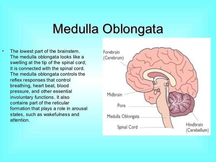
The medulla oblongata, often simply called the medulla, is an elongated section of neural tissue that makes up part of the brainstem. The medulla is anterior to the cerebellum and is the part of the brainstem that connects to the spinal cord. It is continuous with the spinal cord, meaning there is not a clear delineation between the spinal cord and medulla but rather the spinal cord gradually transitions into the medulla.
Don’t Miss: Tylenol Heart Rate
Can You Recover From A Brainstem Injury
A brainstem injury can have severe effects because the brainstem controls so many of your bodys most basic functions. But people do recover from some types of brainstem injuries.
Its important to get care right away if you suspect a brainstem injury. The sooner you get care, the more likely your healthcare providers can reduce the damage. You may need rehabilitation and other special care after a brainstem injury.
What Part Of The Brain Controls Heart Rate Breathing And Blood Pressure
4.2/5breathingheart ratepart of the brainbrainbrain
Herein, what part of the brain controls blood pressure?
The medulla oblongata controls breathing, blood pressure, heart rhythms and swallowing. Messages from the cortex to the spinal cord and nerves that branch from the spinal cord are sent through the pons and the brainstem.
Subsequently, question is, how does the medulla control the heart? Medulla Oblongata: The medulla oblongata is part of the brainstem. The medulla oblongata controls many of the autonomic functions of the body, meaning involuntary actions. Its main functions include regulation of breathing, heart rate, blood pressure, digestion, swallowing, and sneezing.
Also to know is, what part of the brain that connects the brain to the spinal cord and controls automatic functions such as breathing digestion heart rate and blood pressure?
The brain stem sits beneath your cerebrum in front of your cerebellum. It connects the brain to the spinal cord and controls automatic functions such as breathing, digestion, heart rate and blood pressure.
How does the medulla oblongata control blood pressure?
Baroreceptor FunctionThey send impulses to the cardiovascular center to regulate blood pressure. At lower blood pressures, the degree of stretch is lower and the rate of firing is slower. When the cardiovascular center in the medulla oblongata receives this input, it triggers a reflex that maintains homeostasis.
Recommended Reading: Esophageal Palpitations
Function Of The Medulla Oblongata
The medulla oblongata controls autonomic functions and connects the higher levels of the brain to the spinal cord. It is also responsible for regulating several basic functions of the autonomic nervous system, including:
- Respiration: chemoreceptors
- Reflex centers of vomiting, coughing, sneezing, and swallowing
Divisions Of The Reticular Formation
Traditionally, the nuclei are divided into three columns:
Sagittal division reveals more morphological distinctions. The raphe nuclei form a ridge in the middle of the reticular formation, and directly to its periphery, there is a division called the medial reticular formation. The medial reticular formation is large, has long ascending and descending fibers, and is surrounded by the lateral reticular formation. The lateral reticular formation is close to the motor nuclei of the cranial nerves and mostly mediates their function. The raphe nuclei is the place of synthesis of the neurotransmitter serotonin, which plays an important role in mood regulation.
The medial reticular formation and lateral reticular formation are two columns of neuronal nuclei with ill-defined boundaries that send projections through the medulla and into the mesencephalon . The nuclei can be differentiated by function, cell type, and projections of efferent or afferent nerves. The magnocellular red nucleus is involved in motor coordination, and the parvocellular nucleus regulates exhalation.
Cross Section of the Pons: A cross section of the lower part of the pons showing the pontine reticular formation labeled as #9.
Recommended Reading: Coronary Insufficiency Symptoms
