Histology Of The Arteries And Veins
Synonyms:
Large veins have diameters greater than 10 mm. They have some smooth muscle in all three tunics. They have a relatively thin tunica media with only a moderate amount of smooth muscle the tunica externa is the thickest layer and contains longitudinal bundles of smooth muscle.
Large veins include the venae cavae, pulmonary veins, internal jugular veins, and renal veins. Since the pressure of the pulmonary veins cannot be easily measured, the pulmonary capillary wedge pressure is used instead, and the normal range is 2-15 mmHg. The pulmonary arteries have thin distensible walls with less elastic tissue than the systemic arteries. Thus, they have a blood pressure range of 15-30 mmHg systolic, and 4-12 mmHg diastolic.
What Does The Circulatory System Do
The circulatory system is made up of blood vessels that carry blood away from and towards the heart. Arteries carry blood away from the heart and veins carry blood back to the heart.
The circulatory system carries oxygen, nutrients, and to cells, and removes waste products, like carbon dioxide. These roadways travel in one direction only, to keep things going where they should.
Read Also: Does Acid Reflux Cause Heart Palpitations
Right Vs Left Side Of The Heart
The easiest way to understand the blood flow through the heart is to divide the heart into 2 sides.
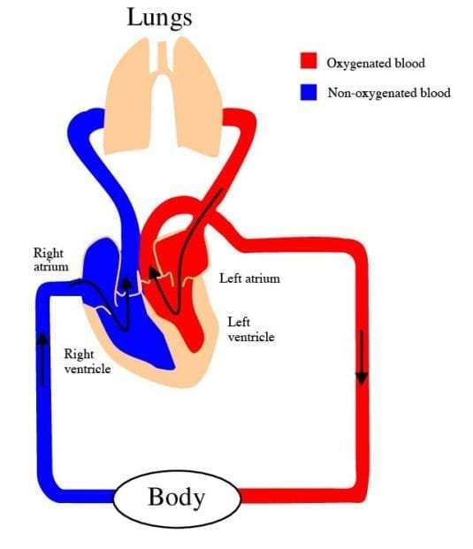
We first have the right side of the heart shown in blue below.
There are 6 main steps or structures in which blood flows through the right side of the heart.
Next, we have the left side of the heart shown in red.
Similar to the right side, there are 6 main steps or structures in which blood flows through the left side of the heart.
Diagram: Blood flow steps through the right and left side of the heart, cardiac anatomy and structures, and cardiac circulation pathway.
Don’t Miss: Flonase And Heart Palpitations
What Are The Parts Of The Circulatory System
Two pathways come from the heart:
- The pulmonary circulation is a short loop from the heart to the lungs and back again.
- The systemic circulation carries blood from the heart to all the other parts of the body and back again.
In pulmonary circulation:
- The pulmonary artery is a big artery that comes from the heart. It splits into two main branches, and brings blood from the heart to the lungs. At the lungs, the blood picks up oxygen and drops off carbon dioxide. The blood then returns to the heart through the pulmonary veins.
In systemic circulation:
How Does A Healthy Heart Work

The heart is part of the circulatory system, which carries blood throughout the body. The heart is made of muscle and works like a pump to keep the blood moving through the blood vessels .
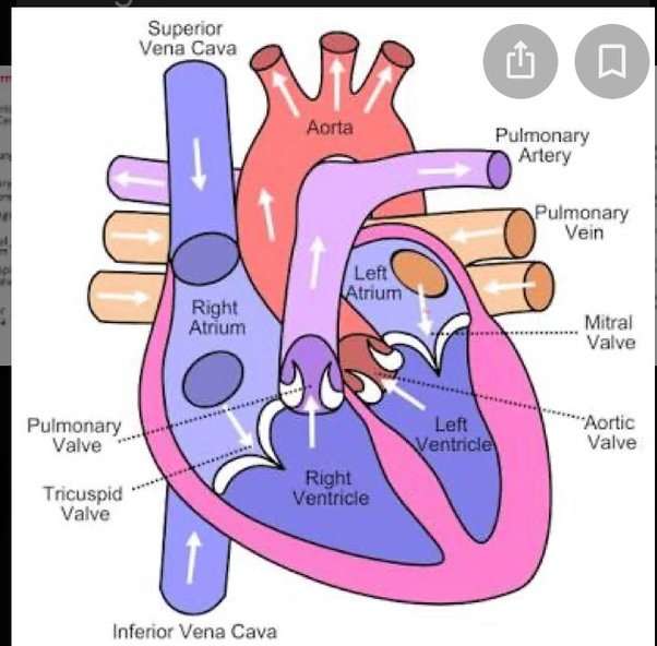
The heart has 4 chambers the right atrium and the left atrium on top and the right and left ventricles on the bottom. The heart is divided by a solid wall called the septum into 2 sides: the right side sends blood to the lungs to get oxygen, while the left side of the heart moves oxygen-rich blood to the rest of the body through the aorta .
Blood enters the heart through the right atrium and moves to the right ventricle, where it then moves through the pulmonary artery to the lungs to pick up oxygen. The newly oxygenated blood then enters the heart through the left atrium and moves to the left ventricle, where it is sent through the aorta to the rest of the body.
There are also 4 valves in the heart, which open and close to allow blood to move through the chambers:
- The aortic valve, located on the left side of the heart, between the aorta and the left ventricle.
- The mitral valve, located between the left ventricle and the left atrium.
- The pulmonary valve, located on the right side of the heart between the right ventricle and the pulmonary artery .
- The tricuspid valve, located on the right side of the heart between the right ventricle and the right atrium.
The exterior of the heart.
Blood vesselsarteries, veins, and capillaries–are also involved in helping blood flow:
Don’t Miss: Left Ventricular Systolic Dysfunction Symptoms
How Your Heart Works
Your heart
The human heart is one of the hardest-working organs in the body.
On average, it beats around 75 times a minute. As the heart beats, it provides pressure so blood can flow to deliver oxygen and important nutrients to tissue all over your body through an extensive network of arteries, and it has return blood flow through a network of veins.
In fact, the heart steadily pumps an average of 2,000 gallons of blood through the body each day.
Your heart is located underneath your sternum and ribcage, and between your two lungs.
The hearts four chambers function as a double-sided pump, with an upper and continuous lower chamber on each side of the heart.
The hearts four chambers are:
- Right atrium. This chamber receives venous oxygen-depleted blood that has already circulated around through the body, not including the lungs, and pumps it into the right ventricle.
- Right ventricle. The right ventricle pumps blood from the right atrium to the pulmonary artery. The pulmonary artery sends the deoxygenated blood to the lungs, where it picks up oxygen in exchange for carbon dioxide.
- Left atrium. This chamber receives oxygenated blood from the pulmonary veins of the lungs and pumps it to the left ventricle.
- Left ventricle. With the thickest muscle mass of all the chambers, the left ventricle is the hardest pumping part of the heart, as it pumps blood that flows to the heart and rest of the body other than the lungs.
What Are The Common Types Of Pediatric Congenital Heart Defects
A septal defect is a hole in the septum, the wall that divides the heart. There are 2 types of septal defects: atrial septal defects are holes in the septum between the left and the right atria ventricular septal defects are holes in the septum between the left and right ventricles. Because of this hole, oxygenated blood mixes with non-oxygenated blood.
A septal defect means that blood flows from one chamber of the heart to the other, instead of taking its normal path. For instance, with an atrial septal defect, blood flows from one atrium to the other, instead of going to the ventricle.
Similarly, with a VSD, the blood flows from the left ventricle to the right ventricle, rather than through its normal path to the aorta and the rest of the body. As a result, blood that has picked up oxygen from the lungs mixes with oxygen-poor blood. This can mean that parts of the body arent getting enough oxygenated blood.
ASDs and VSDs can be small or large. Some ASDs close up on their own as the child grows older. Others may be repaired using catheters or with open heart surgery.
Although some small VSDs may close on their own, some are so large that the left side of the heart is forced to work much harder. If it is not treated, a VSD can lead to heart failure. These defects have to be repaired with open heart surgery.
Valve defects
Another type of defect involves the heart valves. Defective valves may be caused by:
Other types of congenital heart defects
Read Also: What Heart Chamber Pushes Blood Through The Aortic Semilunar Valve
What Are The 4 Types Of Valves
The 4 heart valves are:Tricuspid valve. This valve is located between the right atrium and the right ventricle.Pulmonary valve. The pulmonary valve is located between the right ventricle and the pulmonary artery.Mitral valve. This valve is located between the left atrium and the left ventricle. Aortic valve.
How The Heart Works
The heart is an organ, about the size of a fist. It is made of muscle and pumps blood through the body. Blood is carried through the body in blood vessels, or tubes, called arteries and veins. The process of moving blood through the body is called circulation. Together, the heart and vessels make up the cardiovascular system.
Recommended Reading: Can Ibs Cause Heart Palpitations
The Four Chambers Of The Heart
Your heart has a right and left side separated by a wall called the septum. Each side has a small collecting chamber called an atrium, which leads into a large pumping chamber called a ventricle. There are four chambers: the left atrium and right atrium , and the left ventricle and right ventricle .The right side of your heart collects blood on its return from the rest of our body. The blood entering the right side of your heart is low in oxygen. Your heart pumps the blood from the right side of your heart to your lungs so it can receive more oxygen. Once it has received oxygen, the blood returns directly to the left side of your heart, which then pumps it out again to all parts of your body through an artery called the aorta. Blood pressure refers to the amount of force the pumping blood exerts on arterial walls.
Which Is Side Of The Heart Carries Deoxygenated Blood
The right side of the heart pumps blood to the lungs, and the left side pumps blood to the rest of the body. The blood on the right side is deoxygenated and the blood on the left side is oxygenated. Which side of the heart is deoxygenated blood? the right side of the heart carries deoxygenated blood
Read Also: Can Antihistamines Cause Heart Palpitations
Blood Flow Step By Step
The heart has two upper chambersthe left and right atriumsand two larger lower chambersthe left and right ventricles. A series of valves control blood flow in and out of these chambers.
Electrical impulses, controlled by the cardiac conduction system, make the heart muscle contract and relax, creating the rate and rhythm of your heartbeat. Here are the steps of blood flow through the heart and lungs:
How Do You Identify A Valve

The tag itself should identify the valve, usually by showing the valve number. The type of valve and the system the valve is part of typically are identified. For example, the valve tag might read 200# Main Steam Shut Off. This identifies the valve as the shutoff valve in the 200-pound main steam line.
Read Also: Reflux And Palpitations
What Heart Rate Is Too High
Maximum heart rate and Target Heart Rate
Going beyond your maximum heart rate is not healthy for you. Your maximum heart rate depends on your age. This is how you can calculate it:
- Subtracting your age from the number 220 will give you your maximum heart rate. Suppose your age is 35 years, your maximum heart rate is 185 beats per minute. If your heart rate exceeds 185 beats per minute during exercise, it is dangerous for you.
- Your target heart rate zone is the range of heart rate that you should aim for if you want to become physically fit. It is calculated as 60 to 80 percent of your maximum heart rate.
- Your target heart rate helps you to know if you are exercising at the right intensity.
- It is always better to consult your doctor before starting any vigorous exercise. This is especially important if you have diabetes, heart disease, or you are a smoker. Your doctor might advise you to lower your target heart rate by 50 percent or more.
What Is Left Side Of Heart
Left side of the human heart comprises two chambers, which are left ventricle and left atrium. Furthermore, it has two main heart valves namely aortic valve and bicuspid mitral valves. The left side of the heart receives oxygenated blood from the pulmonary veins and helps to pump it throughout the body cells and organs. Since left ventricle pumps oxygenated blood to all body parts, it needs a heavy force.
Figure 01: Left Side of Heart
Hence, the walls of the left ventricle are thicker than the walls of the right ventricle. Aorta and pulmonary veins connect to the left side of the heart through the left atrium.
When considering the blood flow through the left side of the heart, it occurs as follows.
- Pulmonary veins bring the oxygen-rich blood from the lungs to left
- Then the left atrium contracts and blood flows through the mitral valve to the left ventricle.
- Next, the mitral valve shuts and left ventricles contracts.
- Finally, the blood enters into aortic valve and flows throughout the body.
Read Also: How Does Blood Move Through The Heart
The Valves Are Like Doors To The Chambers Of The Heart
Four valves regulate and support the flow of blood through and out of the heart. The blood can only flow one waylike a car that must always be kept in drive. Each valve is formed by a group of folds, or cusps, that open and close as the heart contracts and dilates. There are two atrioventricular valves, located between the atrium and the ventricle on either side of the heart: The tricuspid valve on the right has three cusps, the mitral valve on the left has two. The other two valves regulate blood flow out of the heart. The aortic valve manages blood flow from the left ventricle into the aorta. The pulmonary valve manages blood flow out of the right ventricle through the pulmonary trunk into the pulmonary arteries.
The Heart Wall Is Composed Of Three Layers
The muscular wall of the heart has three layers. The outermost layer is the epicardium . The epicardium covers the heart, wraps around the roots of the great blood vessels, and adheres the heart wall to a protective sac. The middle layer is the myocardium. This strong muscle tissue powers the hearts pumping action. The innermost layer, the endocardium, lines the interior structures of the heart.
Don’t Miss: Does Tylenol Increase Heart Rate
Electrical Impulses Keep The Beat
The heartâs four chambers pump in an organized manner with the help of electrical impulses that originate in the sinoatrial node . Situated on the wall of the right atrium, this small cluster of specialized cells is the heartâs natural pacemaker, initiating electrical impulses at a normal rate.
The impulse spreads through the walls of the right and left atria, causing them to contract, forcing blood into the ventricles. The impulse then reaches the atrioventricular node, which acts as an electrical bridge for impulses to travel from the atria to the ventricles. From there, a pathway of fibers carries the impulse into the ventricles, which contract and force blood out of the heart.
What Does The Heart Look Like And How Does It Work
- The heart is an amazing organ. It starts beating about 22 days after conception and continuously pumps oxygenated red blood cells and nutrient-rich blood and other compounds like platelets throughout your body to sustain the life of your organs.
- Its pumping power also pushes blood through organs like the lungs to remove waste products like CO2.
- This fist-sized powerhouse beats about 100,000 times per day, pumping five or six quarts of blood each minute, or about 2,000 gallons per day.
- In general, if the heart stops beating, in about 4-6 minutes of no blood flow, brain cells begin to die and after 10 minutes of no blood flow, the brain cells will cease to function and effectively be dead. There are few exceptions to the above.
- The heart works by a regulated series of events that cause this muscular organ to contract and then relax .
- The normal heart has 4 chambers that undergo the squeeze and relax cycle at specific time intervals that are regulated by a normal sequence of electrical signals that arise from specialized tissue.
- In addition, the normal sequence of electrical signals can be sped up or slowed down depending on the needs of the individual, for example, the heart will automatically speed up electrical signals to respond to a person running and will automatically slow down when a person takes a nap.
Read Also: Can Too Much Vitamin D Cause Heart Palpitations
Don’t Miss: Does Flonase Help With Shortness Of Breath
What Part Of The Heart Pumps Blood To The Body
4.2/5side of the heart pumps bloodside of the heartbloodpumpsbodyread here
The right ventricle pumps the oxygen-poor blood to the lungs. The left atrium receives oxygen-rich blood from the lungs and pumps it to the left ventricle. The left ventricle pumps the oxygen-rich blood to the body.
Likewise, how does a heart pump blood? The right side of your heart gets blood from your body and pumps it into your lungs. Oxygen-poor blood flows in through the large veins to the right atrium. Then the blood moves into the right ventricle, which contracts and sends blood out of your heart to pick up oxygen from your lungs.
One may also ask, which part of the heart pumps blood to all parts of the body?
The left side of your heart receives oxygen-rich blood from your lungs and pumps it through your arteries to the rest of your body.
What pumps blood to the lungs?
The heart has a total of four chambers: right atrium, right ventricle, left atrium and left ventricle. The right side of the heart collects oxygen-depleted blood and pumps it to the lungs, through the pulmonary arteries, so that the lungs can refresh the blood with a fresh supply of oxygen.
