Roots Suffixes And Prefixes
Most medical terms are comprised of a root word plus a suffix and/or a prefix . Here are some examples related to the Integumentary System. For more details see Chapter 4: Understanding the Components of Medical Terminology
| component | |
| echocardiogram = sound wave image of the heart. | |
| CYTE- | |
| haematoma – a tumour or swelling filled with blood. | |
| THROMB- | thrombocytopenia = deficiency of thrombocytes in the blood |
| ETHRO- | |
| cerebrovascular = blood vessels of the cerebrum of the brain. | |
| HYPER- | hyperglycaemia = excessive levels of glucose in blood. |
| HYPO- | hypoglycaemia = abnormally low glucose blood levels. |
| -PENIA | neutropenia = low levels of neutrophilic leukocytes. |
| -EMIA |
Chambers And Circulation Through The Heart
The human heart consists of four chambers: The left side and the right side each have one atrium and one ventricle. Each of the upper chambers, the right atrium and the left atrium, acts as a receiving chamber and contracts to push blood into the lower chambers, the right ventricle and the left ventricle. The ventricles serve as the primary pumping chambers of the heart, propelling blood to the lungs or to the rest of the body.
There are two distinct but linked circuits in the human circulation called the pulmonary and systemic circuits. Although both circuits transport blood and everything it carries, we can initially view the circuits from the point of view of gases. The pulmonary circuit transports blood to and from the lungs, where it picks up oxygen and delivers carbon dioxide for exhalation. The systemic circuit transports oxygenated blood to virtually all of the tissues of the body and returns relatively deoxygenated blood and carbon dioxide to the heart to be sent back to the pulmonary circulation.
How Your Heart Works
The heart is a muscular pump which lies at the centre of the human cardiovascular system. The main function of the heart is to circulate blood throughout the body. Oxygen-rich blood flows out of the heart through the aorta, which then branches into smaller arteries carrying blood to all parts of the body. Conversely, oxygen-poor blood is transported back to the heart through a network of veins which culminate in two large veins known as the superior and inferior vena cava respectively.
The heart is divided vertically into two cavities by a muscular wall called the septum. The left cavity pumps blood throughout the body, while the right cavity pumps blood only to the lungs. Each cavity is in turn divided horizontally into two chambers, making a total of four chambers altogether. The two upper chambers are known as the atria, and the two lower chambers as the ventricles. The atria receive blood flowing back to the heart, while the ventricles hold blood that is to be pumped out of the heart.
Inside the right atrium of the heart sits a small bundle of muscle fibres and nerves. This is the sinus or sinoatrial node, which acts as the hearts natural pacemaker.
Also Check: How Does Heart Disease Affect The Skeletal System
Circulatory System: Pulmonary And Systemic Circuits
- B.A., Biology, Emory University
- A.S., Nursing, Chattahoochee Technical College
The circulatory system is a major organ system of the body. This system transports oxygen and nutrients in the blood to all of the cells in the body. In addition to transporting nutrients, the circulatory system also picks up waste products generated by metabolic processes and delivers them to other organs for disposal.
The circulatory system, sometimes called the cardiovascular system, consists of the heart, blood vessels, and blood. The heart provides the “muscle” needed to pump blood throughout the body. Blood vessels are the conduits through which blood is transported and blood contains the valuable nutrients and oxygen that are needed to sustain tissues and organs. The circulatory system circulates blood in two circuits: the pulmonary circuit and systemic circuit.
Valves Maintain Direction Of Blood Flow

As the heart pumps blood, a series of valves open and close tightly. These valves ensure that blood flows in only one direction, preventing backflow.
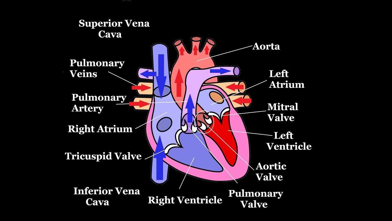
- The tricuspid valve is situated between the right atrium and right ventricle.
- The pulmonary valve is between the right ventricle and the pulmonary artery.
- The mitral valve is between the left atrium and left ventricle.
- The aortic valve is between the left ventricle and the aorta.
Each heart valve, except for the mitral valve, has three flaps that open and close like gates on a fence. The mitral valve has two valve leaflets.
You May Like: Does Tylenol Raise Blood Pressure And Heart Rate
The Exterior Of The Heart:
Below is a picture of the outside of a normal, healthy, human heart.
The illustration shows the front surface of the heart, including the coronary arteries and major blood vessels.
The heart is the muscle in the lower half of the picture. The heart has four chambers. The right and left atria are shown in purple. The right and left ventricles are shown in red.
Connected to the heart are some of the main blood vessels arteries and veins that make up the blood circulatory system.
The ventricle on the right side of the heart pumps blood from the heart to the lungs.
When you breathe air in, oxygen passes from the lungs through blood vessels where its added to the blood. Carbon dioxide, a waste product, is passed from the blood through blood vessels to the lungs and is removed from the body when you breathe air out.
The atrium on the left side of the heart receives oxygen-rich blood from the lungs.
The pumping action of the left ventricle sends this oxygen-rich blood through the aorta to the rest of the body.
S Of Blood Flow Through The Heart
In summary from the video, in 14 steps, blood flows through the heart in the following order: 1) body > 2) inferior/superior vena cava > 3) right atrium > 4) tricuspid valve > 5) right ventricle > 6) pulmonary arteries > 7) lungs > 8) pulmonary veins > 9) left atrium > 10) mitral or bicuspid valve > 11) left ventricle > 12) aortic valve > 13) aorta > 14) body.
Don’t Miss: How Do You Calculate Max Heart Rate
Disorders Of The Heart Valves
When heart valves do not function properly, they are often described as incompetent and result in valvular heart disease, which can range from benign to lethal. Some of these conditions are congenital, that is, the individual was born with the defect, whereas others may be attributed to disease processes or trauma. Some malfunctions are treated with medications, others require surgery, and still others may be mild enough that the condition is merely monitored since treatment might trigger more serious consequences.
Valvular disorders are often caused by carditis, or inflammation of the heart. One common trigger for this inflammation is rheumatic fever, or scarlet fever, an autoimmune response to the presence of a bacterium, Streptococcus pyogenes, normally a disease of childhood.
While any of the heart valves may be involved in valve disorders, mitral regurgitation is the most common, detected in approximately 2 percent of the population, and the pulmonary semilunar valve is the least frequently involved. When a valve malfunctions, the flow of blood to a region will often be disrupted. The resulting inadequate flow of blood to this region will be described in general terms as an insufficiency. The specific type of insufficiency is named for the valve involved: aortic insufficiency, mitral insufficiency, tricuspid insufficiency, or pulmonary insufficiency.
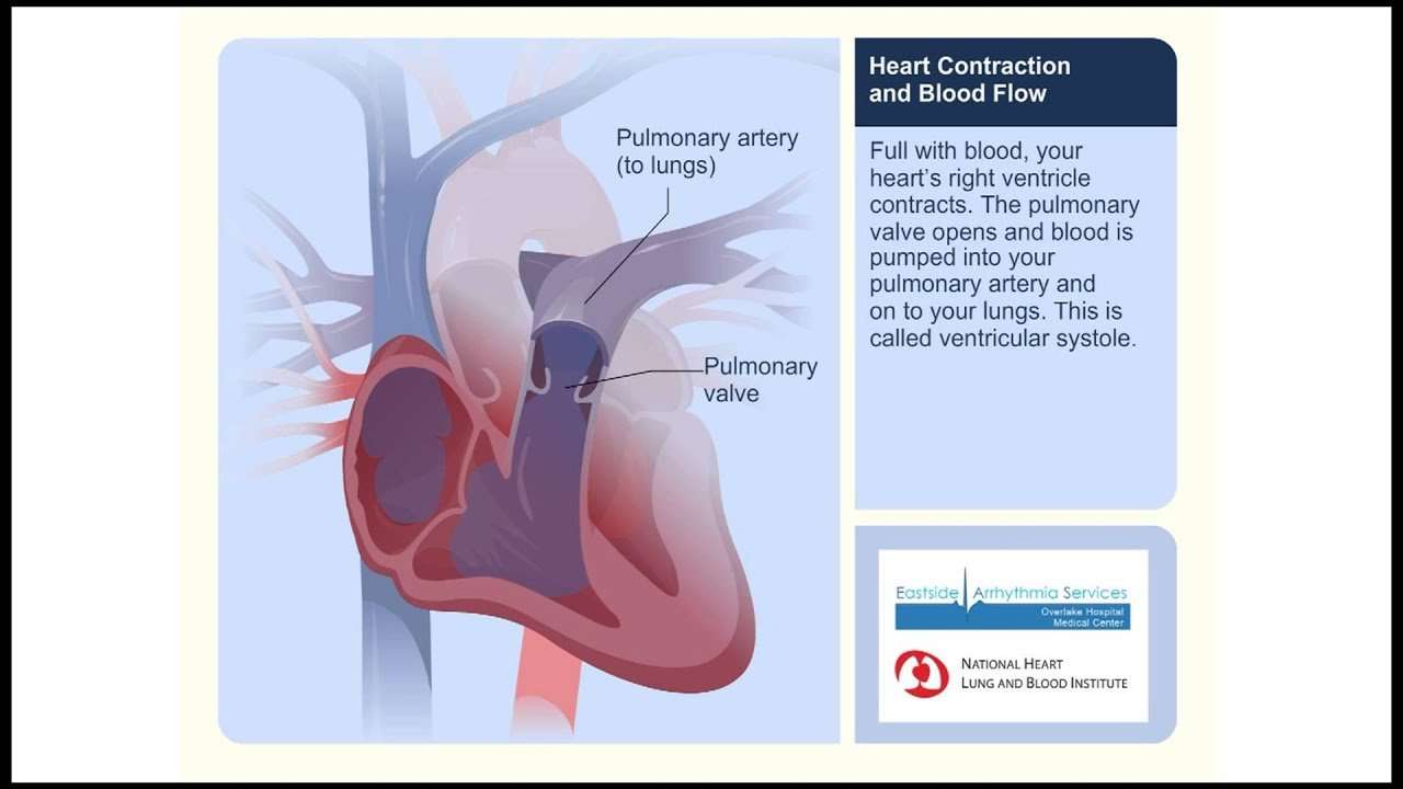
Heart Contraction And Blood Flow
Almost everyone has heard the real or recorded sound of a heartbeat. When the heart beats, it makes a lub-DUB sound. Between the time you hear lub and DUB, blood is pumped through the heart and circulatory system.
A heartbeat may seem like a simple event repeated over and over. A heartbeat actually is a complicated series of very precise and coordinated events that take place inside and around the heart.
Each side of the heart uses an inlet valve to help move blood between the atrium and ventricle.
The tricuspid valve does this between the right atrium and ventricle. The mitral valve does this between the left atrium and ventricle. The lub is the sound of the mitral and tricuspid valves closing.
Each of the hearts ventricles has an outlet valve. The right ventricle uses the pulmonary valve to help move blood into the pulmonary arteries. The left ventricle uses the aortic valve to do the same for the aorta. The DUB is the sound of the aortic and pulmonary valves closing.
Each heartbeat has two basic parts: diastole and atrial and ventricular systole . During diastole, the atria and ventricles of the heart relax and begin to fill with blood.
At the end of diastole, the hearts atria contract and pump blood into the ventricles. The atria then begin to relax. Next, the hearts ventricles contract and pump blood out of the heart.
Don’t Miss: Why Do Av Nodal Cells Not Determine The Heart Rate
Blood Vessels Of The Heart
The blood vessels of the heart include:
- venae cavae deoxygenated blood is delivered to the right atrium by these two veins. One carries blood from the head and upper torso, while the other carries blood from the lower body
- pulmonary arteries deoxygenated blood is pumped by the right ventricle into the pulmonary arteries that link to the lungs
- pulmonary veins the pulmonary veins return oxygenated blood from the lungs to the left atrium of the heart
- aorta this is the largest artery of the body, and it runs the length of the trunk. Oxygenated blood is pumped into the aorta from the left ventricle. The aorta subdivides into various branches that deliver blood to the upper body, trunk and lower body
- coronary arteries like any other organ or tissue, the heart needs oxygen. The coronary arteries that supply the heart are connected directly to the aorta, which carries a rich supply of oxygenated blood
- coronary veins deoxygenated blood from heart muscle is ‘dumped’ by coronary veins directly into the right atrium.
What The Heart And Circulatory System Do
The circulatory system works closely with other systems in our bodies. It supplies oxygen and nutrients to our bodies by working with the respiratory system. At the same time, the circulatory system helps carry waste and carbon dioxide out of the body.
Hormones produced by the endocrine system are also transported through the blood in the circulatory system. As the bodys chemical messengers, hormones transfer information and instructions from one set of cells to another. For example, one of the hormones produced by the heart helps control the kidneys release of salt from the body.
One complete heartbeat makes up a cardiac cycle, which consists of two phases:
In the systemic circulation, blood travels out of the left ventricle, to the aorta, to every organ and tissue in the body, and then back to the right atrium. The arteries, capillaries, and veins of the systemic circulatory system are the channels through which this long journey takes place.
Don’t Miss: How Much Blood Does An Adult Heart Pump Every Day
Heart Diagram Parts Location And Size
Location and size of the heart
Normal heart anatomy and physiology
Normal heart anatomy and physiology need the atria and ventricles to work sequentially, contracting and relaxing to pump blood out of the heart and then to let the chambers refill. When blood leaves each chamber of the heart, it passes through a valve that is designed to prevent backflow of blood. There are four heart valves within the heart:
- Mitral valve between the left atrium and left ventricle
- Tricuspid valve between the right atrium and right ventricle
- Aortic valve between the left ventricle and aorta
- Pulmonic valve between the right ventricle and pulmonary artery
How the heart valves work
Supplying Oxygen To The Hearts Muscle

Like other muscles in the body, your heart needs blood to get oxygen and nutrients. Yourcoronary arteries supply blood to your heart. These arteries branch off from the aorta so that oxygen-rich blood is delivered to your heart as well as the rest of your body.
- The left coronary artery delivers blood to the left side of your heart, including your left atrium and ventricle and the septum between the ventricles.
- The circumflex artery branches off from the left coronary artery to supply blood to part of the left ventricle.
- The left anterior descending artery also branches from the left coronary artery and provides blood to parts of both the right and left ventricles.
- The right coronary artery provides blood to the right atrium and parts of both ventricles.
- The marginal arteries branch from the right coronary artery and provide blood to the surface of the right atrium.
- The posterior descending artery also branches from the right coronary artery and provides blood to the bottom of both ventricles.
Arteries supplying oxygen to the body. The coronary arteries branch off the aorta and supply the heart muscle with oxygen and nutrients. At the top of your aorta, arteries branch off to carry blood to your head and arms. Arteries branching from the middle and lower parts of your aorta supply blood to the rest of your body.
Some conditions can affect normal blood flow through these heart arteries. Examples include:
- The small cardiac vein.
You May Like: Can Flonase Cause Heart Palpitations
What Does The Heart Look Like And How Does It Work
- The heart is an amazing organ. It starts beating about 22 days after conception and continuously pumps oxygenated red blood cells and nutrient-rich blood and other compounds like platelets throughout your body to sustain the life of your organs.
- Its pumping power also pushes blood through organs like the lungs to remove waste products like CO2.
- This fist-sized powerhouse beats about 100,000 times per day, pumping five or six quarts of blood each minute, or about 2,000 gallons per day.
- In general, if the heart stops beating, in about 4-6 minutes of no blood flow, brain cells begin to die and after 10 minutes of no blood flow, the brain cells will cease to function and effectively be dead. There are few exceptions to the above.
- The heart works by a regulated series of events that cause this muscular organ to contract and then relax .
- The normal heart has 4 chambers that undergo the squeeze and relax cycle at specific time intervals that are regulated by a normal sequence of electrical signals that arise from specialized tissue.
- In addition, the normal sequence of electrical signals can be sped up or slowed down depending on the needs of the individual, for example, the heart will automatically speed up electrical signals to respond to a person running and will automatically slow down when a person takes a nap.
Improving Health With Current Research
Learn about the following ways the NHLBI continues to translate current research and science into improved health for people who have heart conditions. Research on this topic is part of the NHLBI’s broader commitment to advancing heart and vascular disease scientific discovery.
Learn about some of the pioneering research contributions we have made over the years that have improved clinical care.
You May Like: Which Of The Following Signs Is Commonly Observed In Patients With Right-sided Heart Failure
Keep Your Heart Happy
Most kids are born with a healthy heart and it’s important to keep yours in good shape. Here are some things that you can do to help keep your heart happy:
- Remember that your heart is a muscle. If you want it to be strong, you need to exercise it. How do you do it? By being active in a way that gets you huffing and puffing, like jumping rope, dancing, or playing basketball. Try to be active every day for at least 30 minutes! An hour would be even better for your heart!
- Eat a variety of healthy foods and avoid foods high in unhealthy fats, such as saturated fats and trans fats .
- Try to eat at least five servings of fruits and vegetables each day.
- Avoid sugary soft drinks and fruit drinks.
- Don’t smoke. It can damage the heart and blood vessels.
Your heart deserves to be loved for all the work it does. It started pumping blood before you were born and will continue pumping throughout your whole life.
Circulatory System: Pulmonary Circuit
The pulmonary circuit is the path of circulation between the heart and the lungs. Blood is pumped to the various places of the body by a process known as the cardiac cycle. Oxygen depleted blood returns from the body to the right atrium of the heart by two large veins called vena cavae. Electrical impulses produced by cardiac conduction cause the heart to contract. As a result, blood in the right atrium is pumped to the right ventricle.
On the next heart beat, the contraction of the right ventricle sends the oxygen-depleted blood to the lungs via the pulmonary artery. This artery branches into left and right pulmonary arteries. In the lungs, carbon dioxide in the blood is exchanged for oxygen at lung alveoli. Alveoli are small air sacs that are coated with a moist film that dissolves air. As a result, gases can diffuse across the thin endothelium of the alveoli sacs.
The now oxygen-rich blood is transported back to the heart by the pulmonary veins. The pulmonary circuit is completed when pulmonary veins return blood to the left atrium of the heart. When the heart contracts again, this blood is pumped from the left atrium to the left ventricle and later to systemic circulation.
Read Also: Fitbit Charge 2 Accuracy Heart Rate
