The Left Side Of The Heart
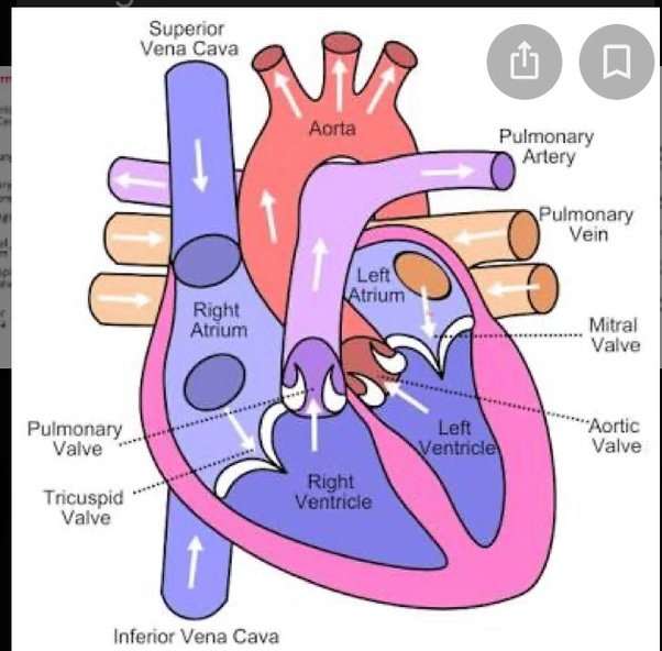
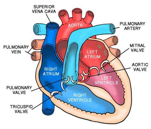
Oxygen-rich blood from the lungs passes through the pulmonary veins . It enters the left atrium and is pumped into the left ventricle. From the left ventricle, the blood is pumped to the rest of the body through the aorta.
Like all of the organs, the heart needs blood rich with oxygen. This oxygen is supplied through the coronary arteries as its pumped out of the hearts left ventricle.
The coronary arteries are located on the hearts surface at the beginning of the aorta. The coronary arteries carry oxygen-rich blood to all parts of the heart.
Blood Supply To The Myocardium
The myocardium of the heart wall is a working muscle that needs a continuous supply of oxygen and nutrients to function efficiently. For this reason, cardiac muscle has an extensive network of blood vessels to bring oxygen to the contracting cells and to remove waste products.
The right and left coronary arteries, branches of the ascending aorta, supply blood to the walls of the myocardium. After blood passes through the capillaries in the myocardium, it enters a system of cardiac veins. Most of the cardiac veins drain into the coronary sinus, which opens into the right atrium.
Left Side Of The Heart
- The pulmonary veins empty oxygen-rich blood from the lungs into the left atrium of the heart.
- As the atrium contracts, blood flows from your left atrium into your left ventricle through the open mitral valve.
- When the ventricle is full, the mitral valve shuts. This prevents blood from flowing backward into the atrium while the ventricle contracts.
- As the ventricle contracts, blood leaves the heart through the aortic valve, into the aorta and to the body.
Also Check: How Accurate Is Fitbit Charge 2 Heart Rate
Pathway Of Blood Through The Heart
While it is convenient to describe the flow of blood through the right side of the heart and then through the left side, it is important to realize that both atria and ventricles contract at the same time. The heart works as two pumps, one on the right and one on the left, working simultaneously. Blood flows from the right atrium to the right ventricle, and then is pumped to the lungs to receive oxygen. From the lungs, the blood flows to the left atrium, then to the left ventricle. From there it is pumped to the systemic circulation.
What Is Left Side Of Heart

Left side of the human heart comprises two chambers, which are left ventricle and left atrium. Furthermore, it has two main heart valves namely aortic valve and bicuspid mitral valves. The left side of the heart receives oxygenated blood from the pulmonary veins and helps to pump it throughout the body cells and organs. Since left ventricle pumps oxygenated blood to all body parts, it needs a heavy force.
Figure 01: Left Side of Heart
Hence, the walls of the left ventricle are thicker than the walls of the right ventricle. Aorta and pulmonary veins connect to the left side of the heart through the left atrium.
When considering the blood flow through the left side of the heart, it occurs as follows.
- Pulmonary veins bring the oxygen-rich blood from the lungs to left
- Then the left atrium contracts and blood flows through the mitral valve to the left ventricle.
- Next, the mitral valve shuts and left ventricles contracts.
- Finally, the blood enters into aortic valve and flows throughout the body.
Recommended Reading: Does Tylenol Increase Heart Rate
What Causes Pediatric Congenital Heart Defects
In most cases, the reasons defects happen are not known, but some connections have been identified:
- Women who get German measles during their first trimester of pregnancy have a higher risk of having a baby with a congenital heart defect.
- The risk may also be higher if the woman has some types of viral infections, is exposed to industrial solvents, takes certain kinds of medications, drinks alcohol, or uses cocaine while pregnant.
- Women who have given birth to a child with a congenital heart defect are at higher risk of giving birth to another child with a heart defect.
Heart defects can also occur along with other types of birth defects.
Electrical Impulses Keep The Beat
The heart’s four chambers pump in an organized manner with the help of electrical impulses that originate in the sinoatrial node . Situated on the wall of the right atrium, this small cluster of specialized cells is the heart’s natural pacemaker, initiating electrical impulses at a normal rate.
The impulse spreads through the walls of the right and left atria, causing them to contract, forcing blood into the ventricles. The impulse then reaches the atrioventricular node, which acts as an electrical bridge for impulses to travel from the atria to the ventricles. From there, a pathway of fibers carries the impulse into the ventricles, which contract and force blood out of the heart.
Also Check: Which Of The Following Signs Is Commonly Observed In Patients With Right-sided Heart Failure
Difference Between Left And Right Side Of Heart
March 6, 2013 Posted by Samanthi
The key difference between left and right side of heart is that the left side of heart comprises of left atrium and left ventricle that have oxygen-rich blood while right side of heart comprises of right atrium and right ventricle that have poor oxygen blood.
The human heart is muscular four-chambered an amazing organ that consists of two ventricles and two atria. It is about the size of fist and situates posterior to the sternum and anterior to the vertebral column in the rib cage. Also, the cardiac muscles make the muscles of the heart, and these muscles contract involuntarily. Furthermore, the main function of the heart is to pump and to circulate blood through the bodys network of blood vessels, which supplies nutrition and oxygen to body cells and removes waste products from the cells.
Since there are two cycle pumps present in humans , the heart can be divided into two parts namely left side and right side, each of which contains one atrium and one ventricle. The muscular part between these two parts is the atrioventricular septum. However, left and right side of heart work together. In simple words, they beat together.
What Does The Heart Look Like And How Does It Work
- The heart is an amazing organ. It starts beating about 22 days after conception and continuously pumps oxygenated red blood cells and nutrient-rich blood and other compounds like platelets throughout your body to sustain the life of your organs.
- Its pumping power also pushes blood through organs like the lungs to remove waste products like CO2.
- This fist-sized powerhouse beats about 100,000 times per day, pumping five or six quarts of blood each minute, or about 2,000 gallons per day.
- In general, if the heart stops beating, in about 4-6 minutes of no blood flow, brain cells begin to die and after 10 minutes of no blood flow, the brain cells will cease to function and effectively be dead. There are few exceptions to the above.
- The heart works by a regulated series of events that cause this muscular organ to contract and then relax .
- The normal heart has 4 chambers that undergo the squeeze and relax cycle at specific time intervals that are regulated by a normal sequence of electrical signals that arise from specialized tissue.
- In addition, the normal sequence of electrical signals can be sped up or slowed down depending on the needs of the individual, for example, the heart will automatically speed up electrical signals to respond to a person running and will automatically slow down when a person takes a nap.
Read Also: Can Too Much Vitamin D Cause Heart Palpitations
Blood Flow Of The Heart Review
Lets now use the 2×2 table we made in the anatomy of the heart post, and this will give us another way to visualize the blood flow through the heart.
Right Side
First, we have the SVC and IVC that carry deoxygenated venous blood from the rest of the body to the right atrium.
Blood will then flow from the right atrium, through the tricuspid valve, and enter the right ventricle.
The deoxygenated blood will then exit the right ventricle, travel through the pulmonary valve, and enter the main pulmonary artery to ultimately be delivered to the lungs to become oxygenated.
Left Side
The oxygenated blood will then travel from the lungs to the left atrium via the pulmonary veins.
Blood will then flow from the left atrium, through the mitral valve, and enter the left ventricle.
The oxygenated blood will then exit the left ventricle, travel through the aortic valve, and enter the aorta to be delivered to the rest of the body.
Diagram: Blood flow through the heart, cardiac circulation pathway steps, and cardiac anatomy and structures. Blue arrows Red arrows .
Now that we have a good understanding of the blood flow through the heart using the cartoon diagrams, we can apply it to a more realistic image of the heart.
The blue arrows represent the flow of deoxygenated blood through the right side of the heart.
The red arrows represent the flow of oxygenated blood through the left side of the heart.
What Is Right Side Of Heart
Right side of the human heart consists of two heart chambers the right ventricle and right atrium. And also, it connects with two heart valves tricuspid valve and pulmonary valve. The right side of the heart receives deoxygenated blood from body organs through superior and inferior vena cava and returns to lungs through the pulmonary artery. The concentration of CO2 and other wastes are higher in the blood which flows through the right side of the heart, hence calls deoxygenated blood. Furthermore, special types of tissues that help to generate nerve impulse can be found only in this side.
Figure 02: Right Side of Heart
When considering the blood flow through the right side of the heart, it occurs as follows.
- Inferior and superior vena cava bring poor oxygen blood to the right atrium.
- Then, through the tricuspid valve, blood flows to the right ventricle.
- Once right ventricle filled with blood, the tricuspid valve shuts and right ventricle contracts.
- Finally, the blood enters the pulmonary valve and flows to the lungs for oxygenation.
You May Like: Does Acid Reflux Cause Heart Palpitations
How Can You Prevent Heart Attacks And Strokes
According to the American Heart Association, no matter what age you are, your heart can benefit from a healthy diet and adequate physical activity. Tthere are numerous specific suggestions about how you can decrease your risk for heart disease. For example:
- Lower cholesterol .
- Lower tryglicerides.
What Does The Circulatory System Do

The circulatory system is made up of blood vessels that carry blood away from and towards the heart. Arteries carry blood away from the heart and veins carry blood back to the heart.
The circulatory system carries oxygen, nutrients, and to cells, and removes waste products, like carbon dioxide. These roadways travel in one direction only, to keep things going where they should.
Read Also: Does Acid Reflux Cause Heart Palpitations
How Does The Heart Beat
The atria and ventricles work together, alternately contracting and relaxing to pump blood through your heart. This is your heartbeat. The electrical system of your heart is the power source that makes this possible.
Your heartbeat is triggered by electrical impulses that travel down a special pathway through your heart.
- The impulse starts in a small bundle of specialized cells called the SA node , located in the right atrium. This node is known as the heart’s natural pacemaker. The electrical activity spreads through the walls of the atria and causes them to contract.
- A cluster of cells in the center of the heart between the atria and ventricles, the AV node is like a gate that slows the electrical signal before it enters the ventricles. This delay gives the atria time to contract before the ventricles do.
- The His-Purkinje network is a pathway of fibers that sends the electrical impulse from the AV node to the muscular walls of the ventricles, causing them to contract.
At rest, a normal heart beats around 50 to 90 times a minute. Exercise, emotions, anemia, an overactive thyroid, fever, and some medications can cause your heart to beat faster, sometimes to well over 100 beats per minute.
Blood Vessels Of The Heart
The blood vessels of the heart include:
- venae cavae deoxygenated blood is delivered to the right atrium by these two veins. One carries blood from the head and upper torso, while the other carries blood from the lower body
- pulmonary arteries deoxygenated blood is pumped by the right ventricle into the pulmonary arteries that link to the lungs
- pulmonary veins the pulmonary veins return oxygenated blood from the lungs to the left atrium of the heart
- aorta this is the largest artery of the body, and it runs the length of the trunk. Oxygenated blood is pumped into the aorta from the left ventricle. The aorta subdivides into various branches that deliver blood to the upper body, trunk and lower body
- coronary arteries like any other organ or tissue, the heart needs oxygen. The coronary arteries that supply the heart are connected directly to the aorta, which carries a rich supply of oxygenated blood
- coronary veins deoxygenated blood from heart muscle is ‘dumped’ by coronary veins directly into the right atrium.
Read Also: How To Calculate Target Heart Rate Zone
How Are Pediatric Congenital Heart Defects Treated
Many children who are born with heart defects do not need treatment. In these cases, the defects are mild or they simply correct on their own .
For children who have a heart defect that must be treated, there are 2 main options: treatment with a catheter, or open heart surgery.
Catheter treatment
Treatment with a catheter is much easier for the child to go through than surgery. Instead of opening the body with an incision as in surgery, the doctor makes a small cut in the skin and inserts a catheter into the body through an artery or vein.
Catheters are used to treat simple heart defects, such as an atrial septal defect. In this procedure, the catheter is moved through a vein until it reaches the septum . There, the catheter places a small device into the septal defect to close it up. The catheter is then removed.
To treat pulmonary valve stenosis, the catheter is equipped with a small balloon that is inflated at the pulmonary valve in order to separate the fused leaflets.
Open heart surgery
In cases where the heart defect cannot be treated with a catheter, the child may need open heart surgery. In these situations, the pediatric heart surgeon opens the chest and operates directly on the heart to repair the defect. This type of treatment is usually done for more serious heart defects.
Heart Anatomy: By The Numbers
1. Superior vena cava: Receives blood from the upper body delivers blood into the right atrium.
2. Inferior vena cava: Receives blood from the lower extremities, pelvis and abdomen, and delivers blood into the right atrium.
3. Right atrium: Receives blood returning to the heart from the superior and inferior vena cava transmits blood to the right ventricle, which pumps blood to the lungs for oxygenation.
4. Tricuspid valve: Allows blood to pass from the right atrium to the right ventricle prevents blood from flowing back into the right atrium as the heart pumps .
5. Right ventricle: Receives blood from the right atrium pumps blood into the pulmonary artery.
6. Pulmonary valve: Allows blood to pass into the pulmonary arteries prevents blood from flowing back into the right ventricle.
7. Pulmonary arteries: Carry oxygen-depleted blood from the heart to the lungs.
8. Pulmonary veins: Deliver oxygen-rich blood from the lungs to the left atrium of the heart.
9. Left atrium: Receives blood returning to the heart from the pulmonary veins.
10. Mitral valve: Allows blood to flow into the left ventricle prevents blood from flowing back into the left atrium.
11. Left ventricle: Receives oxygen-rich blood from the left atrium and pumps blood into the aorta.
12. Aortic valve: Allows blood to pass from the left ventricle to the aorta prevents backflow of blood into the left ventricle.
13. Aorta: Distributes blood throughout the body from the heart.
Don’t Miss: Tylenol Heart Rate
Changes In Oxygen Demand
The heart regulates the amount of vasodilation or vasoconstriction of the coronary arteries based upon the oxygen requirements of the heart. This contributes to the filling difficulties of the coronary arteries.Compression remains the same. Failure of oxygen delivery caused by a decrease in blood flow in front of increased oxygen demand of the heart results in tissue ischemia, a condition of oxygen deficiency. Brief ischemia is associated with intense chest pain, known as angina. Severe ischemia can cause the heart muscle to die from hypoxia, such as during a myocardial infarction. Chronic moderate ischemia causes contraction of the heart to weaken, known as myocardial hibernation.
In addition to metabolism, the coronary circulation possesses unique pharmacologic characteristics. Prominent among these is its reactivity to adrenergic stimulation.
Structure Of The Heart
The heart consists of four chambers separated into two sides. Each side contains an atria which receives blood into the heart and flows it into a ventricle, which pumps the blood out of the heart. The atria and ventricle on each side of the heart are linked together by valves that prevent backflow of blood. The wall that separates the left and right side of the heart is called the septum.
The left heart deals with systemic circulation, while the right heart deals with pulmonary circulation. The left side of the heart receives oxygenated blood from the pulmonary vein and pumps it into the aorta, while the right side of the heart receives deoxygenated blood from the vena cava and pumps it into the pulmonary vein. The pulmonary vein and aorta also have valves connecting them to their respective ventricle.
Don’t Miss: Why Do Av Nodal Cells Not Determine The Heart Rate
