What Are The Radiological Findings Of Congestive Heart Failure
The radiological tests conducted and their findings are as mentioned below:
Chest X-Ray
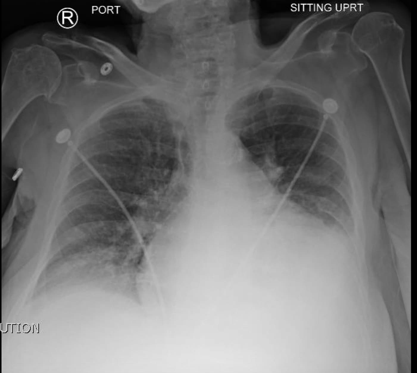
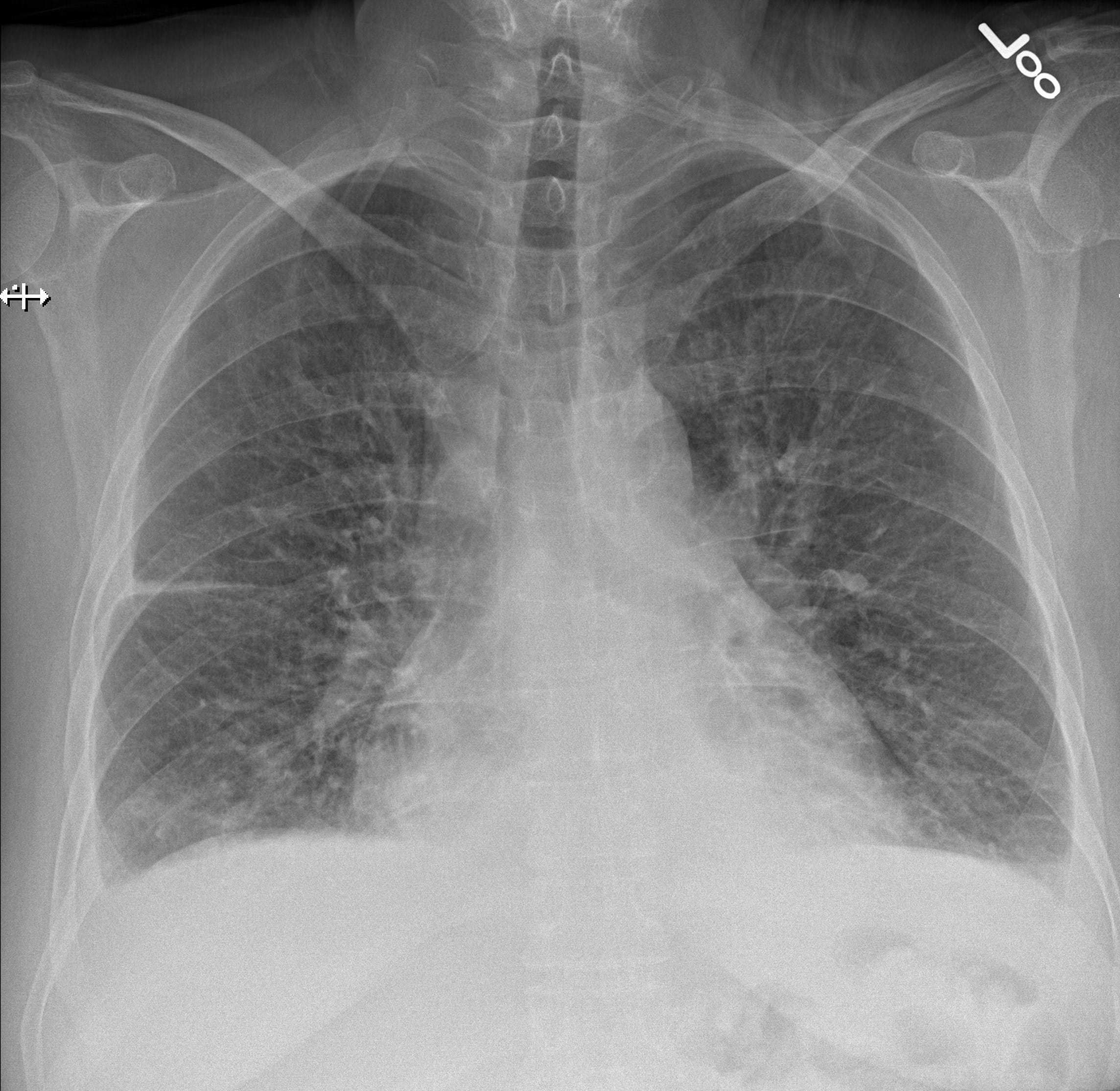
A chest X-ray can be used to view both the heart and lungs. The chest X-ray can assess the size of the heart and the fluid accumulation in the lungs.
Based on the progression of CHF, 3 phases have been described, which are as follows:
– Phase 1: Vascular Phase
-
This represents the first phase of CHF and signifies pulmonary venous hypertension.
-
Cardiomegaly is evident.
-
Prominent upper pulmonary vessels, in contrast to lower blood vessels, are evident in healthy individuals.
-
Hilar level sees an increase in the artery to bronchus ratio, which appears as white round densities.
-
The pulmonary artery is more prominent in diameter than the bronchi .
-
Hilar haziness and fullness: Pulmonary veins are enlarged, and fluid is seen collecting around the vessels.
-
Vascular redistribution is not seen in supine X-rays .
– Phase 2: Interstitial Phase
-
They occur due to interstitial edema and amplified lymphatic drainage.
-
The bronchial wall thickening appears as a white rim around the bronchioles, which appear dark.
-
Thickening of the fissures between the lobes of the lungs.
– Phase 3: Alveolar Phase
Computed Tomography
-
CT is not generally recommended to diagnose heart failure. However, it can reveal any congenital or valvular diseases if present.
-
Thickening of the septal lines will be evident.
-
Ground-glass opacity appearance .
Magnetic Resonance Imaging
Stress Test
Coronary Angiogram
Most Popular Articles
Chest Xray In Congestive Heart Failure
Aka: Chest XRay in Congestive Heart Failure, CHF Chest XRay, Chest XRay in Left Ventricular Failure
II. Pathophysiology: Progression of chronic CHF
III. Imaging: Stage I CHF XRay findings – Vascular phase – pulmonary venous Hypertension
IV. Imaging: Stage II CHF XRay findings – Interstitial phase
Carrus Health Advanced Radiology Department
At Carrus Health, we provide the imaging tests you need for a clinical diagnosis of congestive heart failure. We currently offer CT Scans, X-rays, and ultrasounds to diagnose your condition. Our radiology facility in Sherman, Texas is spacious, clean, and comfortable.
To schedule an appointment, please call Carrus Health at 870-2600. Our friendly staff looks forward to assisting you.
You May Like: Congestive Heart Disease
What Are The Risks Of X
X-rays are monitored and regulated, so you get the minimum amount of radiation exposure needed to produce the image.
For most X-rays, the risk of cancer or defects is very low. Most experts feel that the benefits of appropriate X-ray imaging greatly outweigh any risks.
Young children and babies in the womb are more sensitive to the risks of X-rays. Tell your health care provider if you think you might be pregnant.
What Are The Ways To Diagnose Congestive Heart Failure

Various tests can be used to diagnose the health of the heart. They are described below:
-
Physical Examination and History of the Patient: A detailed physical examination, medical history, family history, and personal history could help ascertain the risk factors and the predisposition for heart failure.
-
Blood Test: Blood tests can determine any risk factors that adversely affect the heart. High serum cholesterol levels could result in blocked coronary arteries. Another blood marker for heart failure is B-type natriuretic peptide . When heart failure occurs, there is a change in blood pressure which prompts the heart to release BNP.
-
Radiological Imaging: Various radiological imaging tests such as X-rays, electrocardiograms, computed tomography , and magnetic resonance imaging are also used to diagnose heart failure.
Also Check: How To Figure Out Target Heart Rate
Use Of Chest Radiography In The Emergency Diagnosis Of Acute Congestive Heart Failure
N MuellerLenkeJ RudezD StaubK LauleKilianT KlimaA P PerruchoudC MuellerKeywords: Copyright
The objective of the present study was to evaluate the diagnostic accuracy of chest radiography in the diagnosis of congestive heart failure in a contemporary cohort of consecutive patients presenting with acute dyspnoea to the emergency department.
Citation Doi & Article Data
Citation:DOI:Bálint BotzRevisions:see full revision historySystems:
- Interstitial pulmonary oedema
Pulmonary interstitial edema represents a form of pulmonary edema resulting from pathological fluid buildup in the interstitial spaces due to increased hydrostatic driving pressure.
Pathology
Interstitial lung edema arises almost exclusively due to an increase of the pulmonary capillary hydrostatic pressure , which occurs most commonly in left sided heart failure, hence it is a key element of cardiogenic lung edema. The increased Pcap leads to an excess filtrate filling the bronchovascular interstitium , and lymphatic distension . Interstitial edema can quickly progress into an alveolar pattern, where the alveolar spaces became flooded too 1.
Don’t Miss: How To Reduce Heart Rate
Assessment Of Myocardial Viability
For patients with angina, known coronary artery disease , previous infarction, and LV dysfunction, a reliable method for assessing the presence, extent, and location of viable myocardium is of considerable clinical importance. It is well established that global or regional ischemic LV dysfunction is not always an irreversible condition. Approximately 25-40% of patients may experience improved function after adequate revascularization.
Two important practical issues must be addressed when patients with presumed ischemic dysfunction are evaluated: assessment of relative regional myocardial uptake of thallium-201 , 99mTc-sestamibi, or 99mTc-tetrofosmin and assessment of the presence of demonstrable ischemia in a myocardial segment with decreased uptake .
How Is A Chest X
A machine beams x-rays that pass through the chest and onto a special film or digital recording plate placed behind your back or to your side, producing a black-and-white image of the organs inside the chest.
Different parts of the body absorb x-rays differently: the more the x-ray is absorbed, the lighter the tissue appears in the final image.
Bone is very dense and absorbs most x-rays, so little of the x-ray reaches the film plate, causing bones to appear white on the final image.
Softer tissue, such as the heart, is less dense and allows more x-rays to pass through to the film plate, causing the heart to appear gray on an x-ray film.
Hollow organs, such as the lungs and the air in them, allow most x-rays to pass through, so they appear black in the final image. If there is fluid buildup in the lungs, more of the x-rays will be blocked, so these areas will appear lighter than normal lungs.
If you are unable to stand, you may be asked to lie on a table with the x-ray beam above you and the film box beneath you.
A chest x-ray usually takes about 15 minutes.
Read Also: How To Slow Your Heart Rate
How Do I Get Ready For A Chest X
-
Your healthcare provider will explain the procedure to you. Ask any questions you have about the procedure.
-
You may be asked to sign a consent form that gives permission to do the procedure. Read the form carefully and ask questions if anything is not clear.
-
You usually do not need to stop eating or drinking before the test. You also usually will not need medicine to help you relax .
-
Tell your healthcare provider if you are pregnant or think you may be pregnant.
-
Wear clothing that you can easily take off. Or wear clothing that lets the radiologist reach your chest.
-
Tell your healthcare provider if you have any body piercings on your chest.
-
Follow any other instructions your healthcare provider gives you to get ready.
How Is Chf Diagnosed
After reporting your symptoms to your doctor, they may refer you to a heart specialist, or cardiologist.
The cardiologist will perform a physical exam, which will involve listening to your heart with a stethoscope to detect abnormal heart rhythms.
To confirm an initial diagnosis, a cardiologist might order certain diagnostic tests to examine your hearts valves, blood vessels, and chambers.
There are a variety of tests used to diagnose heart conditions. Because these tests measure different things, your doctor may recommend a few to get a full picture of your current condition.
You May Like: Does Pregnancy Increase Heart Rate
Dilatation Of Azygos Vein
Dilation of the azygos vein is a sign of increased right atrial pressure and is usually seen when there is also an increase in the width of the vascular pedicle.The diameter of the azygos vein varies according to the positioning.In the standing position a diameter > 7 mm is most likely abnormal and a diameter > 10 mm is definitely abnormal.In a supine patient > 15 mm is abnormal. An increase of 3 mm in comparison to previous films is suggestive of fluid overload. The difference of the azygos diameter on an inspiration film compared to an expiration film is only 1mm. This means that the diameter of the azygos is a valuable tool whether or not there is good inspiration.
How Is Congestive Heart Failure Diagnosed

Congestive heart failure happens when the heart muscles are pumping inefficiently due to fluid buildup around the heart. Its best to get tested, especially if you are at high risk for the condition or have a family history of it. Coronary artery disease, high blood pressure, and a history of heart attacks are all signs that you may be at a higher risk for congestive heart failure. If you smoke, drink excessively, have diabetes, or kidney disease, you are also at a higher risk.
Heart failure doesnt necessarily happen suddenly. It can be chronic, in which case you wont necessarily know whats going on unless you monitor yourself for signs and symptoms. These include:
- Shortness of breath, even when resting
- Reduced ability to exercise, perform regular chores, and move around
- A rapid or irregular heartbeat
- Persistent coughing or wheezing
- Weight gain as a result from fluid retention
If you notice these symptoms, see a doctor immediately. You will need to get examined and tested for the possible underlying cause of your symptoms. So, how is congestive heart failure diagnosed? Here are a few examples of the tests a doctor can perform to diagnose you. You will not need all of them to get diagnosed. Your doctor will determine which combination of tests are best to get accurate results.
You May Like: What Are The Final Stages Of Congestive Heart Failure
What Is Congestive Heart Failure
Congestive heart failure is a progressive disease in which the heart cannot pump blood efficiently. The heart comprises four chambers two atria make up the upper chamber and two ventricles, the lower chambers. The ventricles pump blood from the heart to the rest of the body, and the atria collect the blood circulated from the body. CHF occurs when the ventricles cannot efficiently pump blood from the heart to the rest of the body, resulting in fluid accumulation in the lower extremities, lungs, abdomen, and liver. CHF could be fatal if not diagnosed and treated early. However, proper treatment, lifestyle changes, and medications could prevent any complications and significantly improve the quality of life. Patients with severe conditions may need surgical intervention.
Upper Zone Vessel Enlargement
The upper zone vessels are normally smaller than the lower zone vessels. Prominence of the upper zone vessels such that they are the same size or larger than the lower zone vessels is a sign of increased pulmonary venous pressure.
Hover on/off image to show/hide findings
Tap on/off image to show/hide findings
Signs of heart failure
- Upper zone vessel enlargement a sign of pulmonary venous hypertension
- Airspace shadowing due to alveolar oedema acutely in a peri-hilar distribution
- Blunt costophrenic angles due to pleural effusions
Clinical information
- Rapid onset of shortness of breath
- Atrial fibrillation
- Left ventricular failure with pulmonary oedema
You May Like: By-pass Heart Surgery
What Are The Symptoms Of Congestive Heart Failure
Heart failure can be a long-standing condition or sudden onset . In addition, the patients may exhibit the following symptoms:
-
Shortness of breath, whether at rest or while doing any activity.
-
Swollen ankles, feet, abdomen, or legs.
-
Irregular heartbeat.
-
A sudden increase in body weight.
-
Wheezing or a constant cough with bloody mucus.
-
Bluish discoloration of the skin due to lack of oxygen.
-
Frequent urination at night.
What Are The Causes Of Congestive Heart Failure
Many medical conditions could weaken or damage the heart. They are as mentioned below:
-
Coronary Artery Disease: The arteries supplying the heart may become obstructed entirely or partially, resulting in depletion of oxygen-rich blood to the heart, causing extensive damage to the heart muscles.
-
Heart Attack: When the blood flow to the heart is completely obliterated, it results in a heart attack. A heart attack could also result in scar tissue, thus compromising its function.
-
Cardiomyopathy: It is an acquired or hereditary disorder affecting the heart muscles. It could be due to drug abuse, alcohol consumption, or certain infections.
-
Valvular Disease: The heart has few valves that check the inflow and outflow of blood. Damage to these valves could strain the heart and weaken it.
-
Myocarditis: Inflammation of the muscles of the heart is myocarditis. It is usually caused by a viral infection such as the COVID-19 virus.
-
Congenital Heart Defects: Congenital heart defects refers to heart diseases that are present from birth. If not treated early, they could also result in congestive heart failure.
-
Arrhythmias: Abnormal heartbeat could result in the irregular working of the heart, thus weakening it over time.
-
Risk Factors: Certain risk factors associated with heart failure are diabetes, hypertension, high cholesterol, smoking, alcohol abuse, obesity, and certain medications.
Also Check: What Can I Do To Slow My Heart Rate Down
What Happens During A Chest X
You may have a chest X-ray as an outpatient or as part of your stay in ahospital. The way the test is done may vary depending on your condition andyour healthcare providers practices.
Generally, a chest X-ray follows this process:
You will be asked to remove any clothing, jewelry, or other objects that may get in the way of the test.
You will be given a gown to wear.
You may be asked to lie down, sit, or stand. Your position depends on what images the technologist needs.
For a standing or sitting image, you will stand or sit in front of the X-ray plate. You will be asked to roll your shoulders forward, take in a deep breath, and hold it until the X-ray is made. If you are unable to hold your breath, the technologist will take the picture by watching how you breathe.
You will need to stay still during the X-ray. Moving during the X-ray may affect the quality of the image.
For a side-angle view of the chest, you will be asked to turn to your side and raise your arms above your head. You will be told to take in a deep breath and hold it as the X-ray is made.
The technologist will step behind a special window while the images are being made.
The chest X-ray is not painful. But you may have some discomfort or painfrom moving into different positions if you have had recent surgery or aninjury. The technologist will use all possible comfort measures and do thescan as quickly as possible to minimize any discomfort or pain.
What Are The Risks Of A Chest X
You may want to ask your healthcare provider about the amount of radiationused during the test. Also ask about the risks as they apply to you.
Consider writing down all X-rays you get, including past scans and X-raysfor other health reasons. Show this list to your healthcare provider. Therisks of radiation exposure may be tied to the number of X-rays you haveand the X-ray treatments you have over time.
Tell your healthcare provider if you are pregnant or think you may bepregnant. Radiation exposure during pregnancy may lead to birth defects.
You may have other risks depending on your specific health condition. Talkwith your healthcare provider about any concerns you have before theprocedure.
Also Check: How Long Is Heart Transplant Surgery
Congestive Heart Failure Drugs
There are several medications that can be used to treat CHF, including ACE inhibitors, beta-blockers, and more.
ACE inhibitors
Angiotensin-converting enzyme inhibitors open up narrowed blood vessels to improve blood flow. Vasodilators are another option if you cant tolerate ACE inhibitors.
You may be prescribed one of the following:
voluntary recall of 5 lots of the drug Accupril due to the presence of nitrosamine. Nitrosamine, a known carcinogen with the potential to cause cancer, was found to exist in the drug at levels greater than the Acceptable Daily Intake as determined by the FDA. This recall is specific only to a handful of lot numbers and does not affect all Accupril tablets made by Pfizer. If you take Accupril tablets, talk with your pharmacist or doctor and they will help you determine if your medication has been impacted by the recall.
ACE inhibitors shouldnt be taken with the following medications without consulting a doctor, because they may cause an adverse reaction:
- Potassium-sparing diuretics and potassium supplements. These diuretics can cause potassium buildup in the blood, which may lead to abnormal heart rhythms. Examples include: riamterene , eplerenone , and spironolactone .
- Nonsteroidal anti-inflammatory drugs .NSAIDs such as ibuprofen, aspirin, and naproxen, can cause sodium and water retention. This may reduce the ACE inhibitors effect on your blood pressure.
Beta-blockers
This may be achieved with:
Diuretics
Your doctor may recommend:
